. 2016 May 9:7:120.
doi: 10.3389/fphar.2016.00120. eCollection 2016.

Rifaximin Improves Clostridium difficile Toxin A-Induced Toxicity in Caco-2 Cells by the PXR-Dependent TLR4/MyD88/NF-κB Pathway

Affiliations

Rifaximin Improves Clostridium difficile Toxin A-Induced Toxicity in Caco-2 Cells by the PXR-Dependent TLR4/MyD88/NF-κB Pathway

Giuseppe Esposito et al. Front Pharmacol. .

Abstract

Background: Clostridium difficile infections (CDIs) caused by Clostridium difficile toxin A (TcdA) lead to severe ulceration, inflammation and bleeding of the colon, and are difficult to treat.

Aim: The study aimed to evaluate the effect of rifaximin on TcdA-induced apoptosis in intestinal epithelial cells and investigate the role of PXR in its mechanism of action.

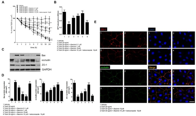
Methods: Caco-2 cells were incubated with TcdA and treated with rifaximin (0.1-10 μM) with or without ketoconazole (10 μM). The transepithelial electrical resistance (TEER) and viability of the treated cells was determined. Also, the expression of zona occludens-1 (ZO-1), toll-like receptor 4 (TLR4), Bcl-2-associated X protein (Bax), transforming growth factor-β-activated kinase-1 (TAK1), myeloid differentiation factor 88 (MyD88), and nuclear factor-kappaB (NF-κB) was determined.

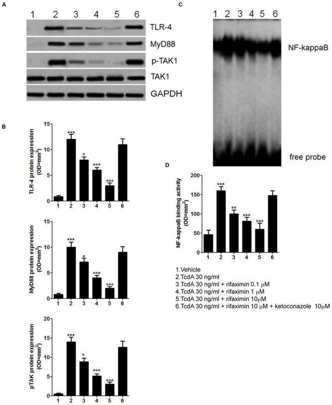
Results: Rifaximin treatment (0.1, 1.0, and 10 μM) caused a significant and concentration-dependent increase in the TEER of Caco-2 cells (360, 480, and 680% vs. TcdA treatment) 24 h after the treatment and improved their viability (61, 79, and 105%). Treatment also concentration-dependently decreased the expression of Bax protein (-29, -65, and -77%) and increased the expression of ZO-1 (25, 54, and 87%) and occludin (71, 114, and 262%) versus TcdA treatment. The expression of TLR4 (-33, -50, and -75%), MyD88 (-29, -60, and -81%) and TAK1 (-37, -63, and -79%) were also reduced with rifaximin versus TcdA treatment. Ketoconazole treatment inhibited these effects.

Conclusion: Rifaximin improved TcdA-induced toxicity in Caco-2 cells by the PXR-dependent TLR4/MyD88/NF-κB pathway mechanism, and may be useful in the treatment of CDIs.

Keywords: Caco-2 cells; Clostridium difficile toxin A; pregnane X receptor; pseudomembranous colitis; rifaximin.


Figures

FIGURE 1
FIGURE 1
Effects of increasing concentrations of rifaximin (0.1, 1.0, and 10 μM) alone and rifaximin plus ketoconazole (10 μM) against TcdA (30 ng/ml) in Caco-2 cells: (A) 24-h time course TEER changes (n = 4); (B) MTT cell viability absorbance at 24 h (n = 5); (C) Immunoreactive bands corresponding to Bax, ZO-1, and occludin expression at 24 h following the TcdA challenge; (D) Relative densitometric analysis of immunoreactive bands (arbitrary units normalized against the expression of the housekeeping GAPDH protein; n = 5), and (E) Immunofluorescent staining showing the effects of TcdA challenge on ZO-1 and occludin co-expression at 24 h. Nuclei were also investigated using DAPI staining (Scale bar = 25 μm). Results are expressed as mean ± SEM of experiments performed in triplicate. ∗∗∗p < 0.001 and ∗∗p < 0.01 vs. vehicle group; ∘∘∘p < 0.001, ∘∘p < 0.01 and °p < 0.05 vs. TcdA group.
FIGURE 2
FIGURE 2
Rifaximin (0.1, 1.0, and 10 μM) down-regulates the TLR4/MyD88/NF-κB pathway by a PXR-dependent mechanism. (A) Immunoblot showing the TLR4, MyD88, and phosphorylated/unphosphorylated TAK1 protein bands, and (B) Relative densitometric analysis of immunoreactive bands (arbitrary units normalized against the expression of the housekeeping GAPDH protein) showing the effects of rifaximin, given alone or in the presence of ketoconazole (10 μM), on the expression of TLR4, MyD88, and pTAK1 in Caco-2 cell line. (C) EMSA analysis showing concentration-dependent inhibition of NF-κB activation by rifaximin, and (D) Relative densitometric analysis of NF-κB bands. Results are expressed as mean ± SEM of n = 5 experiments performed in triplicate. ∗∗∗p < 0.001 vs. vehicle group; ∘∘∘p < 0.001, ∘∘p < 0.01 and °p < 0.05 vs. TcdA group.

Similar articles

Cited by

References

    1. Akira S., Hoshino K. (2003). Myeloid differentiation factor 88-dependent and -independent pathways in toll-like receptor signaling. J. Infect. Dis. 187 S356–S363. 10.1086/374749 - DOI - PubMed
    1. Cash B. D., Lacy B. E., Rao T., Earnest D. L. (2016). Rifaximin and eluxadoline – newly approved treatments for diarrhea-predominant irritable bowel syndrome: what is their role in clinical practice alongside alosetron? Expert Opin. Pharmacother. 17 311–322. 10.1517/14656566.2016.1118052 - DOI - PubMed
    1. Cheng J., Shah Y. M., Ma X., Pang X., Tanaka T., Kodama T., et al. (2010). Therapeutic role of rifaximin in inflammatory bowel disease: clinical implication of human pregnane X receptor activation. J. Pharmacol. Exp. Ther. 335 32–41. 10.1124/jpet.110.170225 - DOI - PMC - PubMed
    1. Dou W., Mukherjee S., Li H., Venkatesh M., Wang H., Kortagere S., et al. (2012). Alleviation of gut inflammation by Cdx2/Pxr pathway in a mouse model of chemical colitis. PLoS ONE 7:e36075 10.1371/journal.pone.0036075 - DOI - PMC - PubMed
    1. Ebigbo A., Messmann H. (2013). Challenges of Clostridium difficile infection. Med. Klin. Intensivmed. Notfmed. 108 624–627. 10.1007/s00063-013-0258-7 - DOI - PubMed