. 2016 May 17:7:124.
doi: 10.3389/fphar.2016.00124. eCollection 2016.

SIRT Is Required for EDP-Mediated Protective Responses toward Hypoxia-Reoxygenation Injury in Cardiac Cells

Affiliations

SIRT Is Required for EDP-Mediated Protective Responses toward Hypoxia-Reoxygenation Injury in Cardiac Cells

Victor Samokhvalov et al. Front Pharmacol. .

Abstract

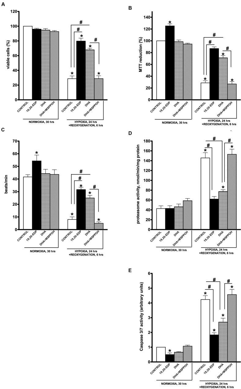
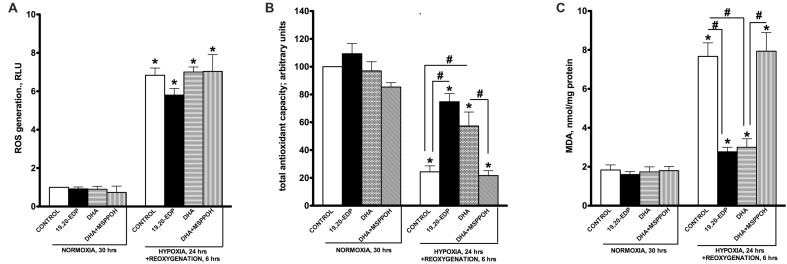
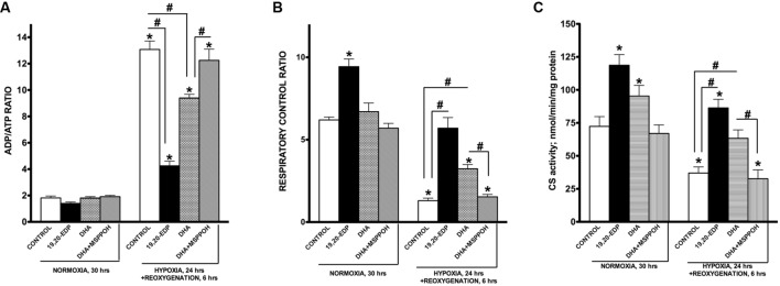
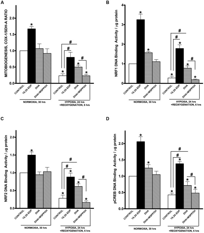
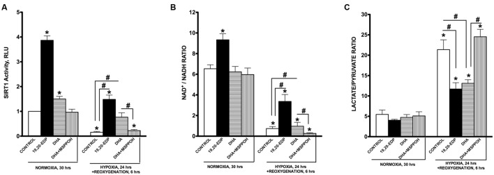
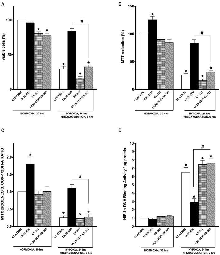
Hypoxia-reoxygenation (H/R) injury is known to cause extensive injury to cardiac myocardium promoting development of cardiac dysfunction. Despite the vast number of studies dedicated to studying H/R injury, the molecular mechanisms behind it are multiple, complex, and remain very poorly understood, which makes development of novel pharmacological agents challenging. Docosahexaenoic acid (DHA, 22:6n3) is an n - 3 polyunsaturated fatty acid obtained from dietary sources, which produces numerous effects including regulation of cell survival and death mechanisms. The beneficial effects of DHA toward the cardiovascular system are well documented but the relative role of DHA or one of its more potent metabolites is unresolved. Emerging evidence indicates that cytochrome P450 (CYP) epoxygenase metabolites of DHA, epoxydocosapentaenoic acids (EDPs), have more potent biological activity than DHA in cardiac cells. In this study we examined whether EDPs protect HL-1 cardiac cells from H/R injury. Our observations demonstrate that treatment with 19,20-EDP protected HL-1 cardiac cells from H/R damage through a mechanism(s) protecting and enhancing mitochondrial quality. EDP treatment increased the relative rates of mitobiogenesis and mitochondrial respiration in control and H/R exposed cardiac cells. The observed EDP protective response toward H/R injury involved SIRT1-dependent pathways.

Keywords: cardiac cells; docosahexaenoic acid; epoxydocosapentaenoic acids; hypoxia–reoxygenation; mitobiogenesis; mitochondrial function.


Figures

FIGURE 1
FIGURE 1
Epoxydocosapentaenoic acids (EDPs) preserve HL-1 cardiac cell viability from hypoxia–reoxygenation (H/R) injury. HL-1 cardiac cells were subjected to either 30 h normoxia or 24 h hypoxia and 6 h reoxygenation in the presence of 19,20-EDP (1 μM), docosahexaenoic acid (DHA; 100 μM) and/or (methylsulfonyl)-2-(2-propynyloxy)-benzenehexanamide (MSPPOH; 50 μM). Treatment of HL-1 cells exposed with DHA or EDPs during H/R injury resulted in (A) preserved cell viability, (B) enhanced mitochondrial activity, and (C) better contractility. Furthermore, both (D) proteasomal, and (E) caspase 3/7 activities were attenuated. Values are represented as mean ± SEM; N = 3 independent experiments; p < 0.05 treatment vs. normoxic control, #p < 0.05 treatment group vs. H/R control or DHA/N-MSPPOH.
FIGURE 2
FIGURE 2
Epoxydocosapentaenoic acids limit H/R-induced cellular oxidative stress responses. HL-1 cardiac cells were subjected to either 30 h normoxia or 24 h hypoxia and 6 h reoxygenation in the presence of 19,20-EDP (1 μM), DHA (100 μM), and/or MSPPOH (50 μM). Treatment of HL-1 cells exposed with DHA or EDPs during H/R injury did not affect generation of ROS (A), while preserved total cellular antioxidant capacity (B) and decreased accumulation of MDA (C). Values are represented as mean ± SEM; N = 3 independent experiments; p < 0.05 treatment vs. normoxic control, #p < 0.05 treatment group vs. H/R control or DHA/MSPPOH.
FIGURE 3
FIGURE 3
EDPs preserve mitochondrial function following H/R injury. HL-1 cardiac cells were subjected to either 30 h normoxia or 24 h hypoxia and 6 h reoxygenation in the presence of 19,20-EDP (1 μM), DHA (100 μM), and/or MSPPOH (50 μM). Treatment of HL-1 cells with DHA or EDPs during H/R injury sustained (A) the intracellular ratio between ADP and ATP, (B) enhanced and protected mitochondrial respiration and finally, (C) limited the drop in citrate synthase (CS) activity (a marker of mitochondrial content) caused by H/R injury. The ratio between basal and ADP-stimulated respiration is presented as respiratory control ratio (RCR). Activity of CS was used as a marker of mitochondrial content. Values are represented as mean ± SEM; N = 3 independent experiments; p < 0.05 treatment vs. normoxic control, #p < 0.05 treatment group vs. H/R control or DHA/MSPPOH.
FIGURE 4
FIGURE 4
EDPs induce mitobiogenesis in HL-1 cardiac cells. HL-1 cardiac cells were subjected to either 30 h normoxia or 24 h hypoxia and 6h reoxygenation in the presence of 19,20-EDP (1 μM), DHA (100 μM), and/or MSPPOH (50 μM). (A) Relative rates of mitobiogenesis were assessed using an ELISA assay detecting simultaneous expression of SDH-A (nDNA-encoded protein) and COX-I (mtDNA-encoded protein) in each well of plated HL-1 cells. The ratio between COX-I and SDH-A expressions represents the relative rate of mitobiogenesis. EDPs increased the relative rates of mitobiogenesis and while preserving the decline following H/R injury. Increased levels of DHA or EDPs in HL-1 cells increased (B) NRF1, (C) NRF2, and (D) pCREB(Ser133) DNA-binding activity. Values are represented as mean ± SEM; N = 3 independent experiments; p < 0.05 treatment vs. normoxic control, #p < 0.05 treatment group vs. H/R control or DHA/MSPPOH.
FIGURE 5
FIGURE 5
EDPs induce SIRT1 activity in HL-1 cardiac cells. HL-1 cardiac cells were subjected to either 30 h normoxia or 24 h hypoxia and 6h reoxygenation in the presence of 19,20-EDP (1 μM), DHA (100 μM), and/or MSPPOH (50 μM). EDPs increased both (A) intracellular SIRT1 enzymatic activity and (B) NAD+/NADH ratios in HL-1 cells. (C) EDPs and DHA prevented the increase in lactate/pyruvate ratio following H/R injury. Values are represented as mean ± SEM; N = 3 independent experiments; p < 0.05 treatment vs. normoxic control, #p < 0.05 treatment group vs. H/R control or DHA/MSPPOH.
FIGURE 6
FIGURE 6
Inhibition of SIRT1 activity abolished 19,20-EDP protective effects against H/R induced cytotoxicity. HL-1 cardiac cells were subjected to either 30 h normoxia or 24 h hypoxia and 6 h reoxygenation in the presence of 19,20-EDP (1 μM) and EX-527 (1 μM) for 24 h. Inhibition of SIRT1 activity blocked DHA and EDP protective effects toward (A) cell viability and (B) mitochondrial activity as assessed by MTT assay. (C) The relative rates of increased mitobiogenesis triggered by EDPs were attenuated by inhibition of SIRT1 activity. (D) EDPs limited HIF-1α DNA-binding activity caused by H/R injury, which were blocked by inhibiting SIRT1 activity. Values are represented as mean ± SEM; N = 3 independent experiments; p < 0.05 treatment vs. normoxic control, #p < 0.05 treatment group vs. H/R control or DHA/MSPPOH.
FIGURE 7
FIGURE 7
Schematic – activation of SIRT1 activity by EDPS is required to exert protective effects. H/R injury overactivates HIF-1α resulting in mitochondrial injury. Subsequently, the compromised mitochondria rapidly promote cell dysfunction and cell death. EDPs act as positive and possibly, selective modulators of SIRT1 activity, through yet to be identified molecular mechanisms, initiating important adaptive responses. SIRT1 can (i) act as a potent suppressor of HIF-1α, (ii) rapidly and potently activate mitobiogenesis, and (iii) selectively eliminate damaged mitochondria via mitophagy. EDP-mediated activation of SIRT1 signaling promotes physiological events that enhance mitochondrial quality control. Thus, preserving a healthy and optimally functioning pool of mitochondria, which protect the cell from LPS-induced toxicity.

Similar articles

Cited by

References

    1. Ahn B. H., Kim H. S., Song S., Lee I. H., Liu J., Vassilopoulos A., et al. (2008). A role for the mitochondrial deacetylase Sirt3 in regulating energy homeostasis. Proc. Natl. Acad. Sci. U.S.A. 105 14447–14452. 10.1073/pnas.0803790105 - DOI - PMC - PubMed
    1. Alcendor R. R., Gao S., Zhai P., Zablocki D., Holle E., Yu X., et al. (2007). Sirt1 regulates aging and resistance to oxidative stress in the heart. Circ. Res. 100 1512–1521. 10.1161/01.RES.0000267723.65696.4a - DOI - PubMed
    1. Ardehali H., Sabbah H. N., Burke M. A., Sarma S., Liu P. P., Cleland J. G., et al. (2012). Targeting myocardial substrate metabolism in heart failure: potential for new therapies. Eur. J. Heart Fail. 14 120–129. 10.1093/eurjhf/hfr173 - DOI - PMC - PubMed
    1. Ayalew-Pervanchon A., Rousseau D., Moreau D., Assayag P., Weill P., Grynberg A. (2007). Long-term effect of dietary {alpha}-linolenic acid or decosahexaenoic acid on incorporation of decosahexaenoic acid in membranes and its influence on rat heart in vivo. Am. J. Physiol. Heart Circ. Physiol. 293 H2296–H2304. 10.1152/ajpheart.00194.2007 - DOI - PubMed
    1. Bayeva M., Gheorghiade M., Ardehali H. (2013). Mitochondria as a therapeutic target in heart failure. J. Am. Coll. Cardiol. 61 599–610. 10.1016/S0735-1097(13)60599-9 - DOI - PMC - PubMed

LinkOut - more resources