. 2016 May 19:7:126.
doi: 10.3389/fphar.2016.00126. eCollection 2016.

EP300 Protects from Light-Induced Retinopathy in Zebrafish

Affiliations

EP300 Protects from Light-Induced Retinopathy in Zebrafish

Reiko Kawase et al. Front Pharmacol. .

Abstract

Exposure of rhodopsin to bright white light can induce photoreceptor cell damage and degeneration. However, a comprehensive understanding of the mechanisms underlying light-induced retinopathy remains elusive. In this study, we performed comparative transcriptome analysis of three rodent models of light-induced retinopathy, and we identified 37 genes that are dysregulated in all three models. Gene ontology analysis revealed that this gene set is significantly associated with a cytokine signaling axis composed of signal transducer and activator of transcription 1 and 3 (STAT1/3), interleukin 6 signal transducer (IL6ST), and oncostatin M receptor (OSMR). Furthermore, the analysis suggested that the histone acetyltransferase EP300 may be a key upstream regulator of the STAT1/3-IL6ST/OSMR axis. To examine the role of EP300 directly, we developed a larval zebrafish model of light-induced retinopathy. Using this model, we demonstrated that pharmacological inhibition of EP300 significantly increased retinal cell apoptosis, decreased photoreceptor cell outer segments, and increased proliferation of putative Müller cells upon exposure to intense light. These results suggest that EP300 may protect photoreceptor cells from light-induced damage and that activation of EP300 may be a novel therapeutic approach for the treatment of retinal degenerative diseases.

Keywords: EP300; STAT3; apoptosis; comparative transcriptome analysis; light-induced retinopathy; systems pharmacology; zebrafish.


Figures

Figure 1
Figure 1
Venn diagram of differentially expressed genes in the three rodent models of light-induced retinopathy. Transcriptome datasets from three different rodent models of light-induced retinopathy (GSE10528, GSE22818, and GSE37773) were downloaded from a public database (GEO). Genes that were differentially expressed in the light-induced retinopathy vs. control groups were identified using a false discovery rate of 10% as the threshold. The number of differentially expressed genes in each transcriptome dataset and the overlap between datasets are shown.
Figure 2
Figure 2
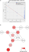
Identification of “STAT transcription factor, coiled coil” as the domain most significantly associated with the DEGs common to the three rodent models of light-induced retinopathy. (A) Scatter plot of domains in InterPro based on the network-based association score (XD score) and the significance of overlap (q-value) using the 37 common DEGs as the input in JEPETTO. The most significant domain was “STAT transcription factor, coiled coil.” (B) The “STAT transcription factor, coiled coil” network identified by JEPETTO. The seven genes with increased expression in the light-induced retinopathy models are shown in red.
Figure 3
Figure 3
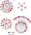
Identification of EP300, STAT1, and STAT3 as the key transcription factors potentially regulating the DEGs common to the three rodent models of light-induced retinopathy. (A–C) The DEGs common to the three models and potentially regulated by EP300 (A), STAT1 (B), and STAT3 (C). (D) The union of the EP300, STAT1, and STAT3 networks and their potential targets.
Figure 4
Figure 4
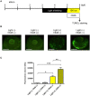
Inhibition of EP300 increases retinal apoptosis in a larval zebrafish model of light-induced retinopathy. (A) Protocol for light-induced retinal damage in larval zebrafish. Zebrafish are shielded from light between 3 and 5 days post-fertilization (dpf) and then exposed to normal conditions (14 h 250 lux/10 h dark) or intense light (13,000 lux) in the presence or absence of 2 μM C646 for 24 h at 27°C. After light exposure, whole-mount TUNEL staining was performed. (B) Representative images of TUNEL staining in the retina of zebrafish exposed to intense light (indicated as light+) or normal light conditions (light-). Scale bars, 100 μm. (C) Quantitative analysis of retinal apoptosis in zebrafish exposed to the conditions shown in (B). *p < 0.01, **p < 0.001, ***p < 0.0001. Data are the mean ± SEM of 13–14 zebrafish/group.
Figure 5
Figure 5
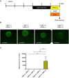
Inhibition of EP300 reduces the photoreceptor cell outer segments in a zebrafish model of light-induced retinopathy. (A) Protocol for light-induced retinal damage in larval zebrafish, as described for Figure 4A. After light exposure, whole-mount immunohistochemical staining with anti-Zpr3 antibody was performed. (B) Representative images of anti-Zpr3 antibody staining of zebrafish exposed to normal or intense light. Scale bars, 100 μm. (C) Quantitative analysis of photoreceptor cell outer segments of zebrafish exposed to the conditions shown in (B). *p < 0.05. Data are the mean ± SEM of n = 24 zebrafish/group for light−/C646- and light−/C646+, n = 17 for light+/C646−, and n = 22 for light+/C646+.
Figure 6
Figure 6
Inhibition of EP300 increases BrdU incorporation in putative Müller cells in the zebrafish model of light-induced retinopathy. (A) Protocol for light-induced retinal damage in larval zebrafish, as described for Figure 4A. After light exposure, whole-mount immunohistochemical staining with anti-BrdU antibody was performed. (B) Representative images of anti-BrdU antibody staining of zebrafish exposed to normal or intense light. Scale bars, 100 μm. (C) Quantitative analysis of BrdU-positive cells in the retinas of zebrafish exposed to the conditions shown in (B). *p < 0.05. Data are the mean ± SEM of n = 8 for all groups.

Similar articles

Cited by

References

    1. Auer T. O., Del Bene F. (2014). CRISPR/Cas9 and TALEN-mediated knock-in approaches in zebrafish. Methods 69, 142–150. 10.1016/j.ymeth.2014.03.027 - DOI - PubMed
    1. Barrett T., Troup D. B., Wilhite S. E., Ledoux P., Rudnev D., Evangelista C., et al. . (2009). NCBI GEO: archive for high-throughput functional genomic data. Nucleic Acids Res. 37, D885–D890. 10.1093/nar/gkn764 - DOI - PMC - PubMed
    1. Bowers E. M., Yan G., Mukherjee C., Orry A., Wang L., Holbert M. A., et al. . (2010). Virtual ligand screening of the p300/CBP histone acetyltransferase: identification of a selective small molecule inhibitor. Chem. Biol. 17, 471–482. 10.1016/j.chembiol.2010.03.006 - DOI - PMC - PubMed
    1. Breitling R., Armengaud P., Amtmann A., Herzyk P. (2004). Rank products: a simple, yet powerful, new method to detect differentially regulated genes in replicated microarray experiments. FEBS Lett. 573, 83–92. 10.1016/j.febslet.2004.07.055 - DOI - PubMed
    1. Bürgi S., Samardzija M., Grimm C. (2009). Endogenous leukemia inhibitory factor protects photoreceptor cells against light-induced degeneration. Mol. Vis. 15, 1631–1637. - PMC - PubMed

LinkOut - more resources