EEG Oscillation Evidences of Enhanced Susceptibility to Emotional Stimuli during Adolescence
- PMID: 27242568
- PMCID: PMC4870281
- DOI: 10.3389/fpsyg.2016.00616
EEG Oscillation Evidences of Enhanced Susceptibility to Emotional Stimuli during Adolescence
Abstract
Background: Our recent event-related potential (ERP) study showed that adolescents are more emotionally sensitive to negative events compared to adults, regardless of the valence strength of the events. The current work aimed to confirm this age-related difference in response to emotional stimuli of diverse intensities by examining Electroencephalography (EEG) oscillatory power in time-frequency analysis.
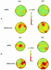
Methods: Time-frequency analyses were performed on the EEG data recorded for highly negative (HN), moderately negative (MN) and Neutral pictures in 20 adolescents and 20 adults during a covert emotional task. The results showed a significant age by emotion interaction effect in the theta and beta oscillatory power during the 500-600 ms post stimulus.
Results: Adolescents showed significantly less pronounced theta synchronization (ERS, 5.5-7.5 Hz) for HN stimuli, and larger beta desynchronization (ERD; 18-20 Hz) for both HN and MN stimuli, in comparison with neutral stimuli. By contrast, adults exhibited no significant emotion effects in theta and beta frequency bands. In addition, the analysis of the alpha spectral power (10.5-12 Hz; 850-950 ms) showed a main effect of emotion, while the emotion by age interaction was not significant. Irrespective of adolescents or adults, HN and MN stimuli elicited enhanced alpha suppression compared to Neutral stimuli, while the alpha power was similar across HN and MN conditions.
Conclusions: These results confirmed prior findings that adolescents are more sensitive to emotionally negative stimuli compared to adults, regardless of emotion intensity, possibly due to the developing prefrontal control system during adolescence.
Keywords: adolescence; event-related desynchronization (ERD); event-related synchronization (ERS); negative stimuli; susceptibility; time-frequency analysis (TFA).
Figures


Similar articles
-
Enhanced brain susceptibility to negative stimuli in adolescents: ERP evidences.Front Behav Neurosci. 2015 Apr 28;9:98. doi: 10.3389/fnbeh.2015.00098. eCollection 2015. Front Behav Neurosci. 2015. PMID: 25972790 Free PMC article.
-
[Reflection of the emotion manifestation in effects of evoked EEG synchronization and desynchronization].Ross Fiziol Zh Im I M Sechenova. 2002 Jun;88(6):790-802. Ross Fiziol Zh Im I M Sechenova. 2002. PMID: 12154576 Russian.
-
Event-related synchronization and desynchronization during affective processing: emergence of valence-related time-dependent hemispheric asymmetries in theta and upper alpha band.Int J Neurosci. 2001;110(3-4):197-219. doi: 10.3109/00207450108986547. Int J Neurosci. 2001. PMID: 11912870
-
Human brain EEG indices of emotions: delineating responses to affective vocalizations by measuring frontal theta event-related synchronization.Neurosci Biobehav Rev. 2011 Oct;35(9):1959-70. doi: 10.1016/j.neubiorev.2011.05.001. Epub 2011 May 10. Neurosci Biobehav Rev. 2011. PMID: 21596060 Review.
-
Event-related EEG/MEG synchronization and desynchronization: basic principles.Clin Neurophysiol. 1999 Nov;110(11):1842-57. doi: 10.1016/s1388-2457(99)00141-8. Clin Neurophysiol. 1999. PMID: 10576479 Review.
Cited by
-
Comparing Neural Correlates of Human Emotions across Multiple Stimulus Presentation Paradigms.Brain Sci. 2021 May 25;11(6):696. doi: 10.3390/brainsci11060696. Brain Sci. 2021. PMID: 34070554 Free PMC article.
-
The regulation of emotions in adolescents: Age differences and emotion-specific patterns.PLoS One. 2018 Jun 7;13(6):e0195501. doi: 10.1371/journal.pone.0195501. eCollection 2018. PLoS One. 2018. PMID: 29879165 Free PMC article. Clinical Trial.
-
Emotional processing of sadness and disgust evoked by disaster scenes.Brain Behav. 2021 Dec;11(12):e2421. doi: 10.1002/brb3.2421. Epub 2021 Nov 22. Brain Behav. 2021. PMID: 34807520 Free PMC article.
-
Neural rhythmic underpinnings of intergroup bias: implications for peace-building attitudes and dialogue.Soc Cogn Affect Neurosci. 2022 Apr 1;17(4):408-420. doi: 10.1093/scan/nsab106. Soc Cogn Affect Neurosci. 2022. PMID: 34519338 Free PMC article.
-
Negative emotional state slows down movement speed: behavioral and neural evidence.PeerJ. 2019 Sep 19;7:e7591. doi: 10.7717/peerj.7591. eCollection 2019. PeerJ. 2019. PMID: 31579576 Free PMC article.
References
-
- Aftanas L., Varlamov A., Pavlov S., Makhnev V., Reva N. (2001). Event-related synchronization and desynchronization during affective processing: emergence of valence related time-dependent hemispheric asymmetries in theta and upper alpha band. Int. J. Neurosci. 110 197–219. 10.3109/00207450108986547 - DOI - PubMed
-
- Aftanas L. I., Reva N. V., Varlamov A. A., Pavlov S. V., Makhnev V. P. (2004). Analysis of evoked EEG synchronization and desynchronization in conditions of emotional activation in humans: temporal and topographic characteristics. Neurosci. Behav. Physiol. 34 859–867. 10.1023/B:NEAB.0000038139.39812.eb - DOI - PubMed
-
- Bai L., Ma H., Huang Y. X. (2005). The development of native chinese affective picture system–a pretest in 46 college students. Chinese Ment. Health J. 19 719–722.
LinkOut - more resources
Full Text Sources
Other Literature Sources

