. 2016 Apr 26:7:588.
doi: 10.3389/fmicb.2016.00588. eCollection 2016.

Photoconversion and Fluorescence Properties of a Red/Green-Type Cyanobacteriochrome AM1_C0023g2 That Binds Not Only Phycocyanobilin But Also Biliverdin

Affiliations

Photoconversion and Fluorescence Properties of a Red/Green-Type Cyanobacteriochrome AM1_C0023g2 That Binds Not Only Phycocyanobilin But Also Biliverdin

Keiji Fushimi et al. Front Microbiol. .

Abstract

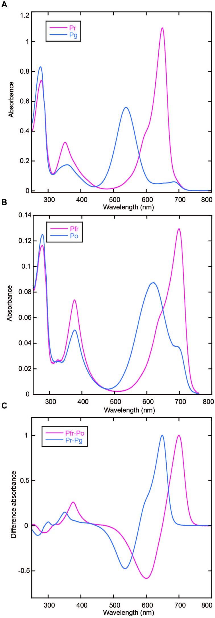
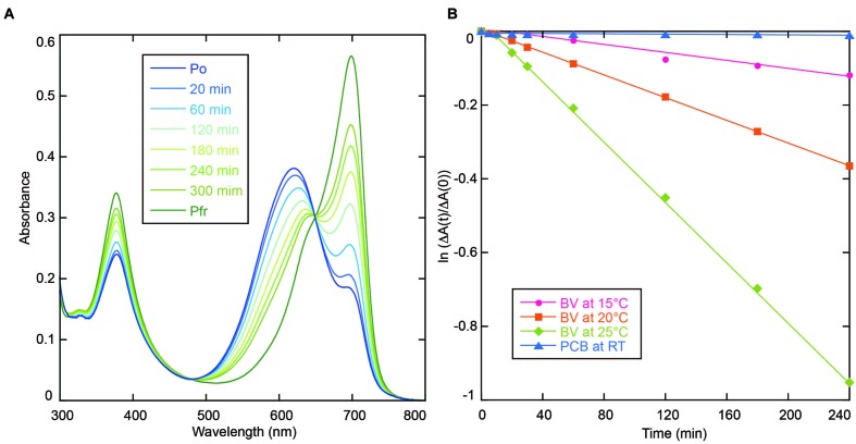
Cyanobacteriochromes (CBCRs) are distantly related to the red/far-red responsive phytochromes. Red/green-type CBCRs are widely distributed among various cyanobacteria. The red/green-type CBCRs covalently bind phycocyanobilin (PCB) and show red/green reversible photoconversion. Recent studies revealed that some red/green-type CBCRs from chlorophyll d-bearing cyanobacterium Acaryochloris marina covalently bind not only PCB but also biliverdin (BV). The BV-binding CBCRs show far-red/orange reversible photoconversion. Here, we identified another CBCR (AM1_C0023g2) from A. marina that also covalently binds not only PCB but also BV with high binding efficiencies, although BV chromophore is unstable in the presence of urea. Replacement of Ser334 with Gly resulted in significant improvement in the yield of the BV-binding holoprotein, thereby ensuring that the mutant protein is a fine platform for future development of optogenetic switches. We also succeeded in detecting near-infrared fluorescence from mammalian cells harboring PCB-binding AM1_C0023g2 whose fluorescence quantum yield is 3.0%. Here the PCB-binding holoprotein is shown as a platform for future development of fluorescent probes.

Keywords: GFP; linear tetrapyrrole; live cell imaging; near-infrared fluorescence; optogenetics.


Figures

FIGURE 1
FIGURE 1
(A) Domain structure of the full-length gene product, AM1_C0023. GAF: cGMP-phosphodiesterase/adenylate cyclase/FhlA domain, HK, Histidine kinase domain; RR, response regulator domain. Expressed second GAF domain, AM1_C0023g2, was highlighted by black solid underline. (B) SDS-PAGE analyses of PCB- and BV-binding AM1_1557g2 and AM1_C0023g2. Upper gel image: Coomassie Brilliant Blue stained gel. Lower gel image: Linear tetrapyrroles covalently bound to CBCR GAF domains were detected by in-gel fluorescence imaging. The Zn2+-staining was performed as previously described (Berkelman and Lagarias, 1986). The gel was directly subjected to fluorescence detection. The gel image was provided with black-and-white inversion for easy detection of fluorescent bands.
FIGURE 2
FIGURE 2
Photoconversion of AM1_C0023g2-PCB and AM1_C0023g2-BV. (A) Absorption spectra of Pr (magenta) and Pg (blue) forms of AM1_C0023g2-PCB. (B) Absorption spectra of Pfr (magenta) and Po (blue) forms of AM1_C0023g2-BV. (C) Difference spectra of PCB- (blue) and BV-binding (magenta) AM1_C0023g2 before and after photoconversion.
FIGURE 3
FIGURE 3
(A) Absorption spectra of AM1_C0023g2-BV recorded during dark reversion at 25°C. (B) Dark reversion kinetics of PCB- (blue triangle for room temperature) and BV-binding (magenta circle for 15°C, orange square for 20°C and green diamond for 25°C) AM1_C0023g2 from metastable Pg and Po forms to thermostable Pr and Pfr forms, respectively.
FIGURE 4
FIGURE 4
Replacement of Ser334 of AM1_C0023g2 with Gly. (A) Photograph of cell pellets harboring apo-AM1_C0023g2, AM1_C0023g2-BV, and S334G-BV. (B) Absorption spectra of the Pfr and Po forms of S334G-BV.
FIGURE 5
FIGURE 5
Acid denaturation of AM1_C0023g2-PCB. (A) Absorption spectra of acid-denatured AM1_C0023g2-PCB Pr. Absorption spectra just after denaturation (Pr, magenta) and after white light illumination (Pr WL, blue). (B) Absorption spectra of acid-denatured AM1_C0023g2-PCB Pg. Absorption spectra just after denaturation (Pg, magenta) and after white light illumination (Pg WL, blue). (C) Pr-minus-Pg difference spectrum (Pr-Pg, magenta) and that during photoconversion of denatured Pg (Pg L-D, blue).
FIGURE 6
FIGURE 6
Acid denaturation of AM1_C0023g2-BV. (A) Absorption spectra of acid-denatured AM1_C0023g2-BV Pfr during dark and light processes. (B) Absorption spectra of acid-denatured AM1_C0023g2-BV Po during dark and light processes. (C) Difference spectrum during dark and light processes of denatured Pfr (magenta), during dark and light processes of denatured Po, and of Pfr-minus-Po just after denaturation (green). (D) Normalized absorption spectra and difference spectrum of acid-denatured AM1_C0023g2-BV Pfr and Po after dark and light processes.
FIGURE 7
FIGURE 7
Denaturation with 1% SDS and in vitro reconstitution. (A) Absorption spectrum of AM1_C0023g2-BV Pfr denatured with 1% SDS. (B) Absorption spectrum of AM1_1557g2-BV Pfr denatured with 1% SDS. (C) Absorption spectra of the Pfr and Po forms of S334G reconstituted with BV in vitro. Photoconversion was repeated twice. First-round Po: Solid blue line, first-round Pfr: Solid magenta line, second-round Po: Dotted cyan line, second-round Pfr: Dotted pink line. Inset: SDS-PAGE of in vitro reconstituted S334G. Left, CBB-stained gel. Right, in-gel fluorescence detection. (D) Pfr-minus-Po difference spectra of S334G reconstituted in vitro and in vivo. In vitro reconstitution: blue (first-round) and magenta (second-round) lines, in vivo reconstitution: black line.
FIGURE 8
FIGURE 8
Fluorescence spectra of AM1_C0023g2-BV Pfr (magenta) and AM1_C0023g2-PCB Pr (blue).
FIGURE 9
FIGURE 9
Expression of GFP-fused AM1_1557g2 and AM1_C0023g2 in mammalian cells. (A) Schematic diagram of plasmid construct of GFP-fused AM1_1557g2 and AM1_C0023g2. Kz, Kozak sequence for efficient translation in mammalian cells; GS, a flexible peptide linker sequence (Gly-Gly-Ser-Gly-Gly); CBCR, AM1_1557g2 or AM1_C0023g2; , stop codon. (B) Representative confocal fluorescence images of GFP-fused AM1_1557g2 with PCB and AM1_C0023g2 with PCB. Left column, GFP-fused AM1_1557g2 with PCB; right column, GFP-fused AM1_C0023g2 with PCB; green images, GFP fluorescence; red images, fluorescence of CBCR with PCB. Scale bars, 20 μm. (C) Relative intensity of fluorescence of CBCR with PCB normalized to GFP fluorescence. Error bars represent standard deviations (n = 16, 16, 16, and 17 for AM1_1557g2 with vehicle, AM1_1557g2 with PCB, AM1_C0023g2 with vehicle and AM1_C0023g2 with PCB, respectively).

References

    1. Anders K., Essen L.-O. (2015). The family of phytochrome-like photoreceptors: diverse, complex and multi-colored, but very useful. Curr. Opin. Struct. Biol. 35 7–16. 10.1016/j.sbi.2015.07.005 - DOI - PubMed
    1. Berkelman T. R., Lagarias J. C. (1986). Visualization of bilin-linked peptides and proteins in polyacrylamide gels. Anal. Biochem. 156 194–201. 10.1016/0003-2697(86)90173-9 - DOI - PubMed
    1. Burgie E. S., Walker J. M., Phillips G. N. J., Vierstra R. D. (2013). A photo-labile thioether linkage to phycoviolobilin provides the foundation for the blue/green photocycles in DXCF-cyanobacteriochromes. Structure 21 88–97. 10.1016/j.str.2012.11.001 - DOI - PubMed
    1. Campbell E. L., Hagen K. D., Chen R., Risser D. D., Ferreira D. P., Meeks J. C. (2015). Genetic Analysis Reveals the Identity of the Photoreceptor for Phototaxis in Hormogonium Filaments of Nostoc punctiforme. J. Bacteriol. 197 782–791. 10.1128/JB.02374-14 - DOI - PMC - PubMed
    1. Chen Y., Zhang J., Luo J., Tu J.-M., Zeng X.-L., Xie J., et al. (2012). Photophysical diversity of two novel cyanobacteriochromes with phycocyanobilin chromophores: photochemistry and dark reversion kinetics. FEBS J. 279 40–54. 10.1111/j.1742-4658.2011.08397.x - DOI - PubMed

LinkOut - more resources