. 2016 May 17:7:184.
doi: 10.3389/fimmu.2016.00184. eCollection 2016.

A T Cell View of the Bone Marrow

Affiliations

A T Cell View of the Bone Marrow

Adriana Bonomo et al. Front Immunol. .

Abstract

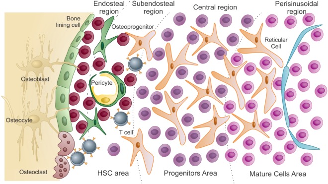
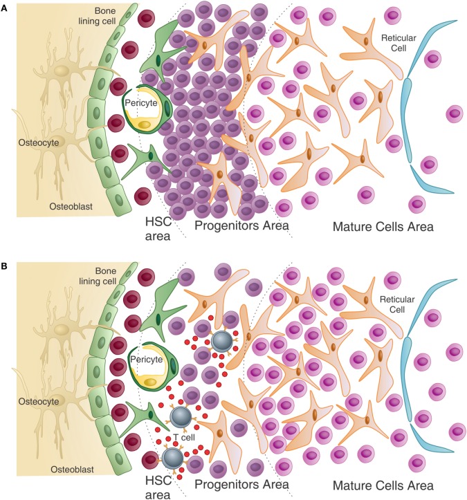
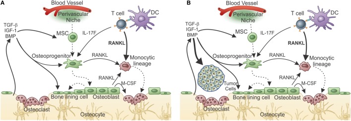
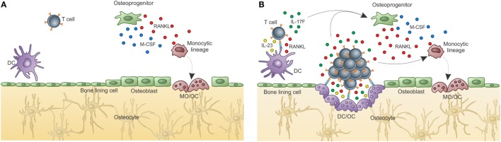
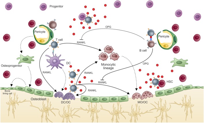
The majority of T cells present in the bone marrow (BM) represent an activated/memory phenotype and most of these, if not all, are circulating T cells. Their lodging in the BM keeps them activated, turning the BM microenvironment into a "memory reservoir." This article will focus on how T cell activation in the BM results in both direct and indirect effects on the hematopoiesis. The hematopoietic stem cell niche will be presented, with its main components and organization, along with the role played by T lymphocytes in basal and pathologic conditions and their effect on the bone remodeling process. Also discussed herein will be how "normal" bone mass peak is achieved only in the presence of an intact adaptive immune system, with T and B cells playing critical roles in this process. Our main hypothesis is that the partnership between T cells and cells of the BM microenvironment orchestrates numerous processes regulating immunity, hematopoiesis, and bone remodeling.

Keywords: B cells; T cells; bone remodeling; hematopoiesis; osteoblast; osteoclast.


Figures

Figure 1
Figure 1
Schematic presentation of the bone marrow microenvironment under “homeostatic” conditions. As previously described by Lambertsen and Weiss (15), HSC area, which harbors hematopoietic stem cells and uncommitted progenitors, comprises both endosteal and subendosteal niches. Committed progenitors and differentiated cells are distributed in the central and perisinusoidal niches, respectively. Quiescent hematopoietic stem cells are in close association with endosteal osteoblasts and bone-lining cells. As HSCs exit quiescence to proliferative states, they migrate and colonize the subendosteal perivascular niche, interacting with both endothelial cells and pericytes. Subendosteal sinusoid-derived pericytes serve as source for new osteoprogenitors, which will differentiate into osteoblasts during bone remodeling (adapted from Journal of Cellular Biochemistry, 2014, Cordeiro-Spinetti et al. with permission from John Wiley & Sons).
Figure 2
Figure 2
T cells help normal hematopoiesis – (A) in the absence of T cells, or in the presence of non-activated T cells (not represented), immature progenitors cells, particularly of the myeloid lineage, accumulate in the bone marrow resulting in peripheral granulopenia. (B) When activated T cells are present, they can see antigen in several hematopoietic cells and release growth factors, in return. These cytokines can directly and indirectly affect maturation of myeloid progenitors. Also, T cell cytokines modulate the stroma, which in turn can modulate HSC maintenance and myeloid differentiation. The number of HSC is also higher than in the T-less environment (adapted from Journal of Cellular Biochemistry, 2014, CordeiroSpinetti et al. with permission from John Wiley & Sons).
Figure 3
Figure 3
Adaptive immunity participates in physiological bone remodeling – (A) bone remodeling relies on osteoclasts (OC), which in physiological situations differentiate from OC precursors of the monocytic lineage after signaling through RANK in its surface and RANKL produced by osteoprogenitor cells. As the bone is resorbed, growth factors and other molecules stored in the extracellular matrix are released, including TGF-β that stimulates osteoblastogenesis from MSC, which reside in the perivascular niche (as in the representation) or in the endosteal niche (not represented). As a consequence, new osteoblasts are formed resulting in bone formation. OPG, a decoy receptor for RANKL, is produced by osteoblasts and participate modulating the bone remodeling system. (B) When activated T cells are present, they activate B cells in a CD40–CD40L-dependent manner with OPG production in large quantities by B cells. This leads to bone accumulation as the RANK–RANKL axis is disrupted by the high amounts of OPG produced by B cells. This is what defines the “normal” bone mass in physiological situation, in SPF condition. Dashed lines represent cellular differentiation; filled line represents cytokine action.
Figure 4
Figure 4
T-cell-derived RANKL prepares the bone pre-metastatic niche. (A) After responding T cells meet tumor antigen and get activated, they migrate to the bone marrow where, after encountering antigen, they release cytokines. In the case of a metastatic tumor, responding T cells produce IL-17 and RANKL. Although IL-17F from T cells can indirectly influences osteoclastogenesis, T-cell-derived RANKL is the central cytokine stimulating osteoclastogenesis and bone consumption resulting in pre-metastatic niche organization with growth factor release from the resorbed matrix. (B) The growth factor-rich environment will nourishes the arriving tumor cells allowing metastatic colonization. In the absence of T-cell-derived RANKL, metastatization into the bone does not take place.
Figure 5
Figure 5
Osteoclasts are optimal antigen-presenting cells when derived from DCs, but not from BM progenitors. Osteoclasts are usually derived from bone marrow progenitors or monocytic precursors (OC), but can also derive from dendritic cells under certain conditions (OC–DC). (A) Classical “constitutive” OC. OC are not good antigen presenters, cannot activate T cells, and do not maintain T cell activation in the BM. Thus, OCs do not contribute to T cell activation in the BM. (B) Activation induced OC–DC. Under the influence of RANKL among other cytokines, OC–DCs are induced. If OC–DCs are as good as fresh DCs in presenting antigen to T cells and if they provide IL-23, a positive loop will be built, which can amplify the osteoclastogenic potential of T cell stimulation. In this context, the bone-resorbing activity of both DC–OC and classical OC will result in decreased bone mass (represented by the light color of the matrix). Whether both OCs dwells in the same resorbing pit, or are segregated into separate subniches, as represented in B, is unknown.
Figure 6
Figure 6
T cell regulation of bone marrow environment. T cell participation in the BM environment promotes what we know as “normal hematopoiesis” and “normal bone mass.” In hematopoiesis, T cell release of hematopoietic cytokines after activation allows terminal differentiation of hematopoietic progenitors/precursors, which will compose the “normal” peripheral pool. “Normal” bone density is also dependent on T cell activation, which in a normal state activates B cells to produce OPG, balancing bone resorption by OCs and mineral deposition by OBs. If T cells are highly activated or are turned into an osteoclastogenic phenotype, excessive bone resorption can lead to osteolytic disease. Moreover, some of the cytokines produced by T cells that can interfere with the bone remodeling system can also affect hematopoiesis, such as IL-17F.

References

    1. Mazo IB, Honczarenko M, Leung H, Cavanagh LL, Bonasio R, Weninger W, et al. Bone marrow is a major reservoir and site of recruitment for central memory CD8+ T cells. Immunity (2005) 22:259–70. 10.1016/j.immuni.2005.01.008 - DOI - PubMed
    1. Sabbagh L, Snell LM, Watts TH. TNF family ligands define niches for T cell memory. Trends Immunol (2007) 28:333–9. 10.1016/j.it.2007.06.001 - DOI - PubMed
    1. Di Rosa F, Pabst R. The bone marrow: a nest for migratory memory T cells. Trends Immunol (2005) 26:360–6. 10.1016/j.it.2005.04.011 - DOI - PubMed
    1. Godin I, Cumano A. The hare and the tortoise: an embryonic haematopoietic race. Nat Rev Immunol (2002) 2:593–604. 10.1038/nri857 - DOI - PubMed
    1. Ikuta K, Weissman IL. Evidence that hematopoietic stem cells express mouse c-kit but do not depend on steel factor for their generation. Proc Natl Acad Sci U S A (1992) 89:1502–6. 10.1073/pnas.89.4.1502 - DOI - PMC - PubMed