

Applications of 2-deoxy-2-fluoro-D-glucose (FDG) in Plant Imaging: Past, Present, and Future
- PMID: 27242806
- PMCID: PMC4860506
- DOI: 10.3389/fpls.2016.00483
Applications of 2-deoxy-2-fluoro-D-glucose (FDG) in Plant Imaging: Past, Present, and Future
Abstract
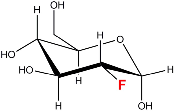
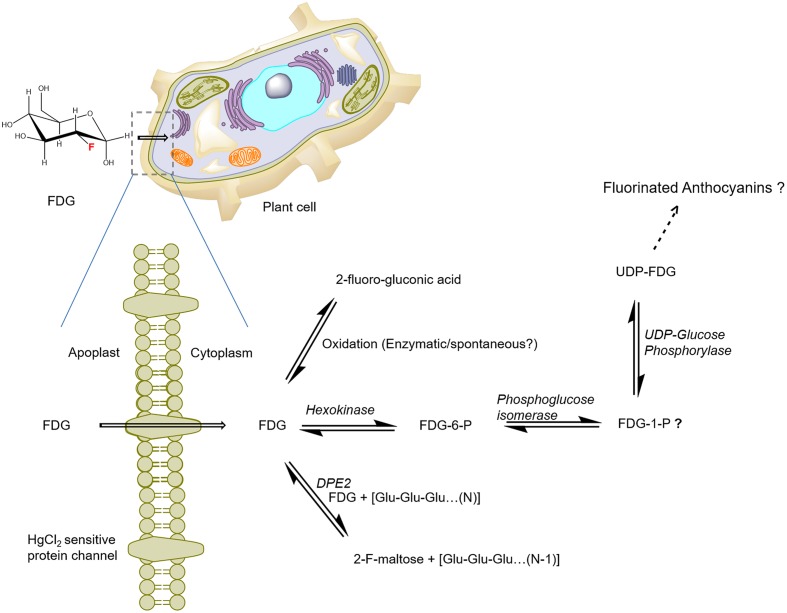
The aim of this review article is to explore and establish the current status of 2-deoxy-2-fluoro-D-glucose (FDG) applications in plant imaging. In the present article, we review the previous literature on its experimental merits to formulate a consistent and inclusive picture of FDG applications in plant-imaging research. 2-deoxy-2-fluoro-D-glucose is a [(18)F]fluorine-labeled glucose analog in which C-2 hydroxyl group has been replaced by a positron-emitting [(18)F] radioisotope. As FDG is a positron-emitting radiotracer, it could be used in in vivo imaging studies. FDG mimics glucose chemically and structurally. Its uptake and distribution are found to be similar to those of glucose in animal models. FDG is commonly used as a radiotracer for glucose in medical diagnostics and in vivo animal imaging studies but rarely in plant imaging. Tsuji et al. (2002) first reported FDG uptake and distribution in tomato plants. Later, Hattori et al. (2008) described FDG translocation in intact sorghum plants and suggested that it could be used as a tracer for photoassimilate translocation in plants. These findings raised interest among other plant scientists, which has resulted in a recent surge of articles involving the use of FDG as a tracer in plants. There have been seven studies describing FDG-imaging applications in plants. These studies describe FDG applications ranging from monitoring radiotracer translocation to analyzing solute transport, root uptake, photoassimilate tracing, carbon allocation, and glycoside biosynthesis. Fatangare et al. (2015) recently characterized FDG metabolism in plants; such knowledge is crucial to understanding and validating the application of FDG in plant imaging research. Recent FDG studies significantly advance our understanding of FDG translocation and metabolism in plants but also raise new questions. Here, we take a look at all the previous results to form a comprehensive picture of FDG translocation, metabolism, and applications in plants. In conclusion, we summarize current knowledge, discuss possible implications and limitations of previous studies, point to open questions in the field, and comment on the outlook for FDG applications in plant imaging.
Keywords: 2-deoxy-2-fluoro-D-glucose; F-sucrose; FDG metabolism; PET; carbon allocation; in vivo imaging; photoassimilate translocation.
Figures
Similar articles
-
Comparing 2-[18F]fluoro-2-deoxy-D-glucose and [68Ga]gallium-citrate translocation in Arabidopsis thaliana.Nucl Med Biol. 2014 Oct;41(9):737-43. doi: 10.1016/j.nucmedbio.2014.05.143. Epub 2014 Jun 9. Nucl Med Biol. 2014. PMID: 25037754
-
2-Deoxy-2-fluoro-d-glucose metabolism in Arabidopsis thaliana.Front Plant Sci. 2015 Nov 3;6:935. doi: 10.3389/fpls.2015.00935. eCollection 2015. Front Plant Sci. 2015. PMID: 26579178 Free PMC article.
-
Incorporation and translocation of 2-deoxy-2-[(18)F]fluoro-D-glucose in Sorghum bicolor (L.) Moench monitored using a planar positron imaging system.Planta. 2008 Apr;227(5):1181-6. doi: 10.1007/s00425-008-0701-9. Epub 2008 Feb 14. Planta. 2008. PMID: 18273639
-
[18F]-Fluoro-2-deoxy-d-glucose-folate.2012 Oct 1 [updated 2012 Oct 31]. In: Molecular Imaging and Contrast Agent Database (MICAD) [Internet]. Bethesda (MD): National Center for Biotechnology Information (US); 2004–2013. 2012 Oct 1 [updated 2012 Oct 31]. In: Molecular Imaging and Contrast Agent Database (MICAD) [Internet]. Bethesda (MD): National Center for Biotechnology Information (US); 2004–2013. PMID: 23136681 Free Books & Documents. Review.
-
2-Deoxy-2-[18F]fluorosorbitol.2008 Jan 14 [updated 2008 Feb 6]. In: Molecular Imaging and Contrast Agent Database (MICAD) [Internet]. Bethesda (MD): National Center for Biotechnology Information (US); 2004–2013. 2008 Jan 14 [updated 2008 Feb 6]. In: Molecular Imaging and Contrast Agent Database (MICAD) [Internet]. Bethesda (MD): National Center for Biotechnology Information (US); 2004–2013. PMID: 20641861 Free Books & Documents. Review.
Cited by
-
Positron Emission Tomography After Ischemic Brain Injury: Current Challenges and Future Developments.Transl Stroke Res. 2020 Aug;11(4):628-642. doi: 10.1007/s12975-019-00765-0. Epub 2020 Jan 14. Transl Stroke Res. 2020. PMID: 31939060 Free PMC article. Review.
-
ID-Checker Technology for the Highly Selective Macroscale Delivery of Anticancer Agents to the Cancer Cells.J Med Chem. 2022 Oct 13;65(19):12883-12894. doi: 10.1021/acs.jmedchem.2c00646. Epub 2022 Oct 4. J Med Chem. 2022. PMID: 36194724 Free PMC article.
-
From the Outside in: An Overview of Positron Imaging of Plant and Soil Processes.Mol Imaging. 2020 Jan-Dec;19:1536012120966405. doi: 10.1177/1536012120966405. Mol Imaging. 2020. PMID: 33119419 Free PMC article. Review.
-
2-NBDG Uptake in Gossypium hirsutum in vitro ovules: exploring tissue-specific accumulation and its impact on hexokinase-mediated glycolysis regulation.Front Plant Sci. 2023 Sep 25;14:1242150. doi: 10.3389/fpls.2023.1242150. eCollection 2023. Front Plant Sci. 2023. PMID: 37818315 Free PMC article.
-
Guide to Plant-PET Imaging Using 11CO2.Front Plant Sci. 2021 Jun 2;12:602550. doi: 10.3389/fpls.2021.602550. eCollection 2021. Front Plant Sci. 2021. PMID: 34149742 Free PMC article.
References
-
- Alavi A., Reivich M., Jones S. C., Greenberg J. H., Wolf A. P. (1982). “Functional imaging of the brain with positron emission tomography,” in Nuclear Medicine Annual 1982, eds Freeman L. M., Weissmann H. S. (New York, NY: Raven Press; ), 319–372.
-
- Alexoff D. L., Dewey S. L., Vaska P., Krishnamoorthy S., Ferrieri R., Schueller M., et al. (2011). PET imaging of thin objects: measuring the effects of positron range and partial-volume averaging in the leaf of Nicotiana tabacum. Nucl. Med. Biol. 38 191–200. 10.1016/j.nucmedbio.2010.08.004 - DOI - PubMed
-
- Avril N. (2004). GLUT1 expression in tissue and 18F-FDG uptake. J. Nucl. Med. 45 930–932. - PubMed
Publication types
LinkOut - more resources
Full Text Sources
Other Literature Sources
Research Materials
Miscellaneous