. 2016:2016:8043983.
doi: 10.1155/2016/8043983. Epub 2016 May 8.

Liposomal Systems as Nanocarriers for the Antiviral Agent Ivermectin

Affiliations

Liposomal Systems as Nanocarriers for the Antiviral Agent Ivermectin

Romina Croci et al. Int J Biomater. 2016.

Abstract

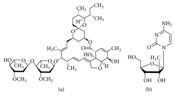
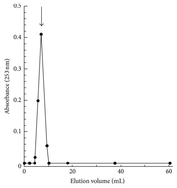
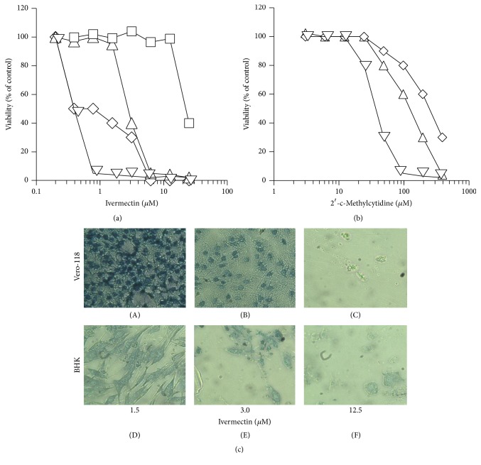
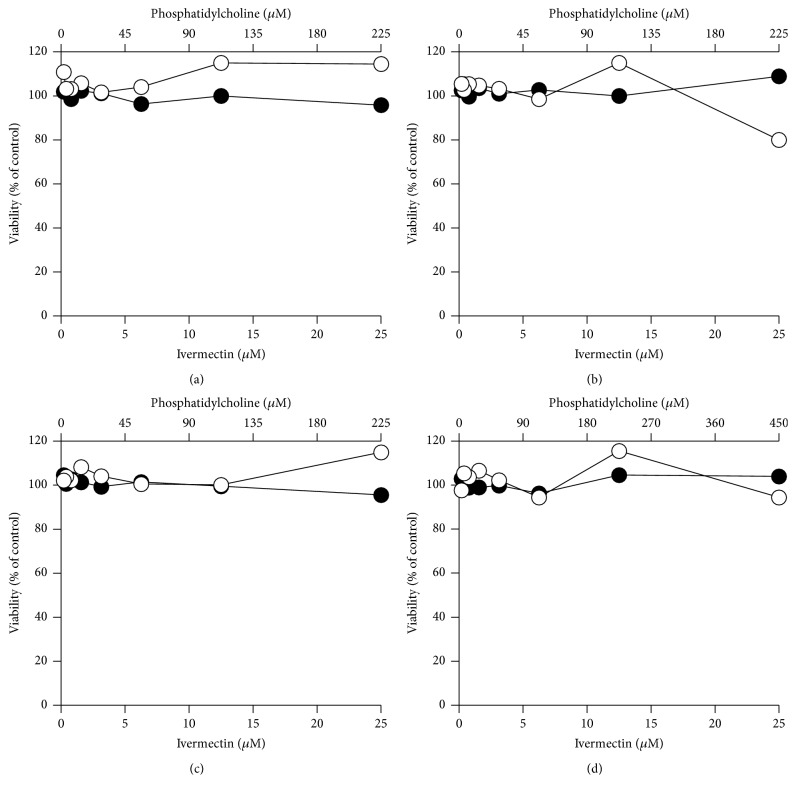
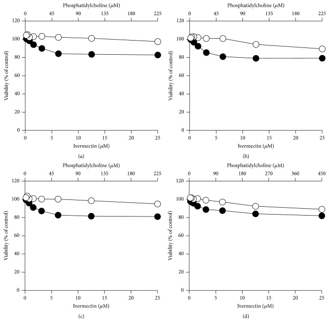
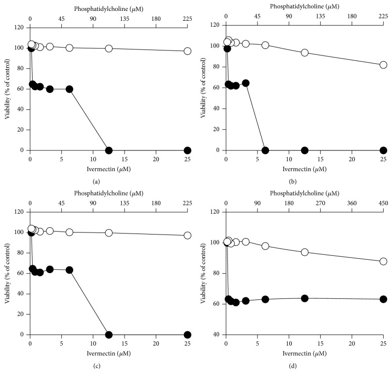
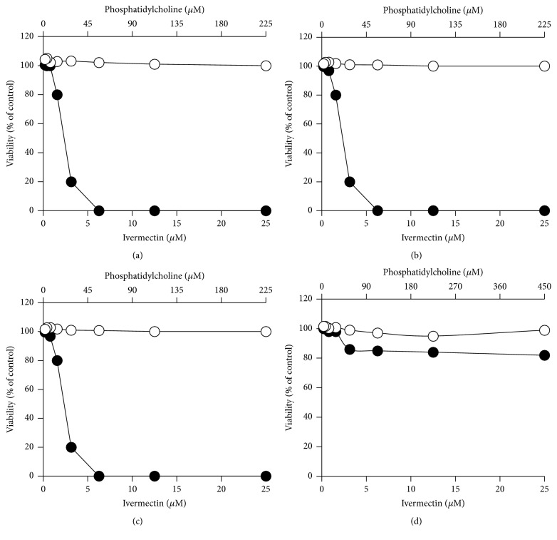
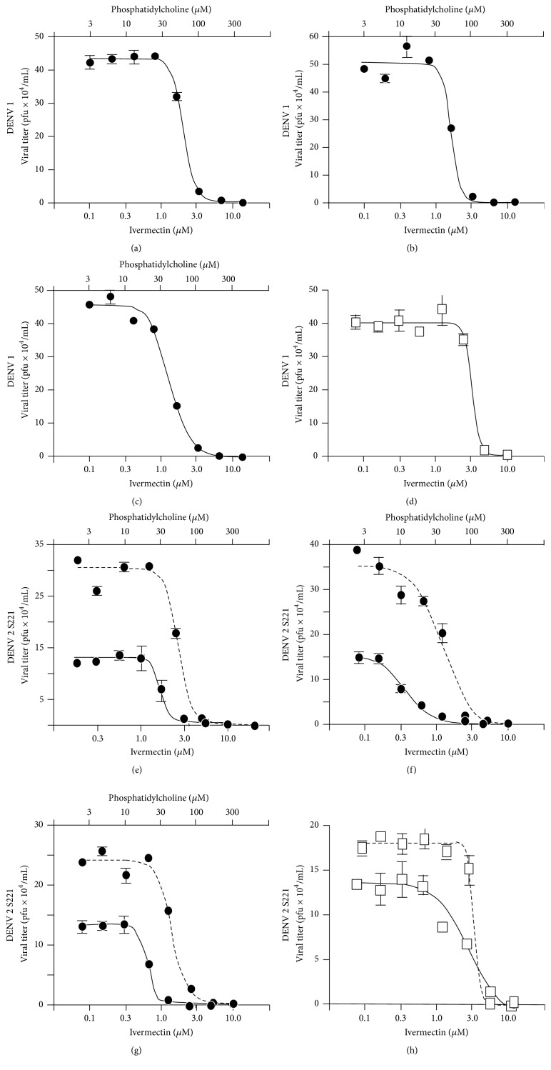
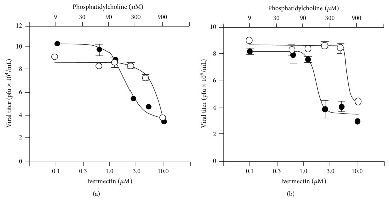
RNA virus infections can lead to the onset of severe diseases such as fever with haemorrhage, multiorgan failure, and mortality. The emergence and reemergence of RNA viruses continue to pose a significant public health threat worldwide with particular attention to the increasing incidence of flaviviruses, among others Dengue, West Nile Virus, and Yellow Fever viruses. Development of new and potent antivirals is thus urgently needed. Ivermectin, an already known antihelminthic drug, has shown potent effects in vitro on Flavivirus helicase, with EC50 values in the subnanomolar range for Yellow Fever and submicromolar EC50 for Dengue Fever, Japanese encephalitis, and tick-borne encephalitis viruses. However ivermectin is hampered in its application by pharmacokinetic problems (little solubility and high cytotoxicity). To overcome such problems we engineered different compositions of liposomes as ivermectin carriers characterizing and testing them on several cell lines for cytotoxicity. The engineered liposomes were less cytotoxic than ivermectin alone and they showed a significant increase of the antiviral activity in all the Dengue stains tested (1, 2, and S221). In the current study ivermectin is confirmed to be an effective potential antiviral and liposomes, as drug carriers, are shown to modulate the drug activity. All together the results represent a promising starting point for future improvement of ivermectin as antiviral and its delivery.


Figures

Figure 1
Figure 1
Chemical structure of ivermectin (a) and 2′-c-methylcytidine (b).
Figure 2
Figure 2
Effect of ivermectin content on the dimensional characteristics of different liposomal formulations (#PC9-Ch1-ive0.1, #PC9-Ch1-ive0.25, #PC9-Ch1-ive0.5, and #PC9-Ch1-ive1.0 in (a); #PC9-Br1-ive0.1, #PC9-Br1-ive0.25, #PC9-Br1-ive0.5, and #PC9-Br1-ive1.0 in (b)). Empty liposomes composed of PC/cholesterol (a) and PC/DDAB (b). Data refers to Z-average (filled circles, left y-axis), mean by number (open squares, right y-axis), and mean by volume (open circles, right y-axis). The arrows indicate the y-axis relative to the arrowed lines.
Figure 3
Figure 3
Cryo-TEM analysis of liposomal formulations, namely, #PC9-Ch1 (a), #PC9-Ch1-ive1.0 (b), #PC9-Br1 (c), and #PC9-Br1-ive1.0 (d). Bar corresponds to 700 nm for (a) and (b) and 100 nm for (c) and (d). For identification and chemical composition of liposomal formulation, see Table 1. In the insets the macroscopic appearance of the formulation is also reported.
Figure 4
Figure 4
Effect of PC content and liposomal composition on mean diameters ((a), (b)) and size distribution ((c)–(f)) of the indicated ivermectin formulations. Bars in (a) and (b) correspond to Z-average (filled bars), mean by number (open bars), and mean by volume (striped bars). Curves in (c) and (e) correspond to #PC3-Ch1-ive0.1 (plain lines) and #PC9-Ch1-ive1.0 (dashed lines) liposomes; curves in (d) and (f) correspond to #PC3-Br1-ive0.1 (plain lines) and #PC9-Br1-ive1.0 (dashed lines) liposomes.
Figure 5
Figure 5
Elution profile of #PC9-Ch1-ive1.0 liposomes on Sepharose 4B gel-filtration column (length: 10 cm, diameter: 1 cm, and flow rate: 160 μL/min). The amount of ivermectin associated with liposomes was determined from the optical density at 253 nm. The solid arrows indicate void volume fractions, including liposome-entrapped ivermectin.
Figure 6
Figure 6
Effect of free ivermectin on the viability of BHK (diamonds), Vero-118 (upward triangles), Vero-F (squares), and RAW (downward triangles) cell lines (a). For comparison, in (b), the experiments are reported, conducted using 2′-c-methylcytidine, employed as reference antiviral compound, on BHK (diamonds), Vero-118 (upward triangles), and RAW (downward triangles) cell lines. (c) Optical microphotograph of Vero-118 ((A), (B), and (C)) and BHK ((D), (E), and (F)) cells treated with the indicated concentration of ivermectin. Data represent the average of 2 experiments.
Figure 7
Figure 7
In vitro cytotoxicity effect of ivermectin formulated in liposomes (closed circles) on Vero-F cells. For comparison the same experiments were performed using empty liposomal formulations (open circles). The following liposomal formulations were tested: #PC9-Ch1 and #PC9-Ch1-ive1.0 (a), #PC9-Br1 and #PC9-Br1-ive1.0 (b), #PC9-Br0.5 and #PC9-Br0.5-ive1.0 (c), and #PC18-Br1 and #PC18-Br1-ive1.0 (d). The phosphatidylcholine concentration (top x-axis) is related to empty liposomes while the ivermectin concentration (bottom x-axis) is related to ivermectin liposomes. Details on the identification codes and liposomal compositions are included in Table 1. Data represent the average of 2 determinations.
Figure 8
Figure 8
In vitro cytotoxicity effect of ivermectin formulated in liposomes (closed circles) on Vero-118 cells. For comparison the same experiments were performed using empty liposomal formulations (open circles). The following liposomal formulations were tested: #PC9-Ch1 and #PC9-Ch1-ive1.0 (a), #PC9-Br1 and #PC9-Br1-ive1.0 (b), #PC9-Br0.5 and #PC9-Br0.5-ive1.0 (c), and #PC18-Br1 and #PC18-Br1-ive1.0 (d). The phosphatidylcholine concentration (top x-axis) is related to empty liposomes while the ivermectin concentration (bottom x-axis) is related to ivermectin liposomes. Details on the identification codes and liposomal compositions are included in Table 1. Data represent the average of 2 determinations.
Figure 9
Figure 9
In vitro cytotoxicity effect of ivermectin formulated in liposomes (closed circles) on BHK cells. For comparison the same experiments were performed using empty liposomal formulations (open circles). The following liposomal formulations were tested: #PC9-Ch1 and #PC9-Ch1-ive1.0 (a), #PC9-Br1 and #PC9-Br1-ive1.0 (b), #PC9-Br0.5 and #PC9-Br0.5-ive1.0 (c), and #PC18-Br1 and #PC18-Br1-ive1.0 (d). The phosphatidylcholine concentration (top x-axis) is related to empty liposomes while the ivermectin concentration (bottom x-axis) is related to ivermectin liposomes. Details on the identification codes and liposomal compositions are included in Table 1. Data represent the average of 2 determinations.
Figure 10
Figure 10
In vitro cytotoxicity effect of ivermectin formulated in liposomes (closed circles) on RAW cells. For comparison the same experiments were performed using empty liposomal formulations (open circles). The following liposomal formulations were tested: #PC9-Ch1 and #PC9-Ch1-ive1.0 (a), #PC9-Br1 and #PC9-Br1-ive1.0 (b), #PC9-Br0.5 and #PC9-Br0.5-ive1.0 (c), and #PC18-Br1 and #PC18-Br1-ive1.0 (d). The phosphatidylcholine concentration (top x-axis) is related to empty liposomes while the ivermectin concentration (bottom x-axis) is related to ivermectin liposomes. Details on the identification codes and liposomal compositions are included in Table 1. Data represent the average of 2 determinations.
Figure 11
Figure 11
In vitro antiviral effect of ivermectin formulated in liposomes on Huh-7 cells infected with DENV 1. Ivermectin (d) and the following liposomal formulations were tested: #PC3-Ch1-ive0.1 (a), #PC3-Cl1-ive0.1 (b), and #PC3-Br1-ive0.1 (c). In vitro antiviral effect of ivermectin formulated in liposomes on Huh-7 cells infected with DENV 2 (plain lines) and DENV 2 mouse adapted S221 strain (dashed lines). The following liposomal formulations were tested: #PC3-Ch1-ive0.1 (e), #PC3-Cl1-ive0.1 (f), and #PC3-Br1-ive0.1 (g). For comparison, data relative to the free ivermectin are reported in (h). The related empty liposomes do not have any effect on the cells. The phosphatidylcholine concentration (top x-axis) is related to empty liposomes while the ivermectin concentration (bottom x-axis) is related to ivermectin liposomes. Details on the identification codes and liposomal compositions are included in Table 1. Details on the EC50 values are in Table 3. Data represent the average of 2 determinations ± SD.
Figure 12
Figure 12
In vitro antiviral effect of ivermectin formulated in liposomes (closed circles) on Huh-7 cells infected with DENV 2. The following liposomal formulations were tested: #PC9-Ch1-ive1.0 (a) and #PC9-Br1-ive1.0 (b). For comparison, the infected cells were treated with the empty liposomal formulations (open circles). The phosphatidylcholine concentration (top x-axis) is related to empty liposomes while the ivermectin concentration (bottom x-axis) is related to ivermectin liposomes. Details on the identification codes and liposomal compositions are included in Table 1. Details on the EC50 values are in Table 3. Data represent the average of 2 determinations ± SD.

References

    1. Vasilakis N., Weaver S. C. The history and evolution of human dengue emergence. Advances in Virus Research. 2008;72:1–76. - PubMed
    1. Guzman M. G., Kouri G. Dengue haemorrhagic fever integral hypothesis: confirming observations, 1987–2007. Transactions of the Royal Society of Tropical Medicine and Hygiene. 2008;102(6):522–523. doi: 10.1016/j.trstmh.2008.03.001. - DOI - PubMed
    1. Kuhn R. J., Zhang W., Rossmann M. G., et al. Structure of dengue virus: implications for flavivirus organization, maturation, and fusion. Cell. 2002;108(5):717–725. doi: 10.1016/s0092-8674(02)00660-8. - DOI - PMC - PubMed
    1. Lindenbach B. D., Rice C. M. Molecular biology of flaviviruses. Advances in Virus Research. 2003;59:23–61. doi: 10.1016/S0065-3527(03)59002-9. - DOI - PubMed
    1. Bollati M., Alvarez K., Assenberg R., et al. Structure and functionality in flavivirus NS-proteins: perspectives for drug design. Antiviral Research. 2010;87(2):125–148. doi: 10.1016/j.antiviral.2009.11.009. - DOI - PMC - PubMed

LinkOut - more resources