

Imaging Evidence for Cerebral Hyperperfusion Syndrome after Intravenous Tissue Plasminogen Activator for Acute Ischemic Stroke
- PMID: 27242938
- PMCID: PMC4868889
- DOI: 10.1155/2016/8725494
Imaging Evidence for Cerebral Hyperperfusion Syndrome after Intravenous Tissue Plasminogen Activator for Acute Ischemic Stroke
Abstract
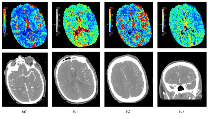
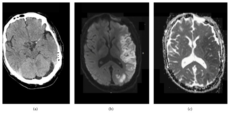
Background. Cerebral hyperperfusion syndrome (CHS), a rare complication after cerebral revascularization, is a well-described phenomenon after carotid endarterectomy or carotid artery stenting. However, the imaging evidence of CHS after intravenous tissue plasminogen activator (iv tPA) for acute ischemic stroke (AIS) has not been reported. Case Report. Four patients were determined to have manifestations of CHS with clinical deterioration after treatment with iv tPA, including one patient who developed seizure, one patient who had a deviation of the eyes toward lesion with worsened mental status, and two patients who developed worsened hemiparesis. In all four patients, postthrombolysis head CT examinations were negative for hemorrhage; CT angiogram showed patent cervical and intracranial arterial vasculature; CT perfusion imaging revealed hyperperfusion with increased relative cerebral blood flow and relative cerebral blood volume and decreased mean transit time along with decreased time to peak in the clinically related artery territory. Vascular dilation was also noted in three of these four cases. Conclusions. CHS should be considered in patients with clinical deterioration after iv tPA and imaging negative for hemorrhage. Cerebral angiogram and perfusion studies can be useful in diagnosing CHS thereby helping with further management.
Figures
Similar articles
-
Cerebral Hyperperfusion Syndrome After Carotid Revascularization and Acute Ischemic Stroke.Curr Pain Headache Rep. 2018 Mar 19;22(4):24. doi: 10.1007/s11916-018-0678-4. Curr Pain Headache Rep. 2018. PMID: 29556806 Review.
-
Cerebral Hyperperfusion Syndrome After Endovascular Reperfusion Therapy in a Patient with Acute Internal Carotid Artery and Middle Cerebral Artery Occlusions.World Neurosurg. 2018 Feb;110:145-151. doi: 10.1016/j.wneu.2017.11.023. Epub 2017 Nov 14. World Neurosurg. 2018. PMID: 29146434
-
Hyperperfusion syndrome after MCA embolectomy - a rare complication?Am J Case Rep. 2013 Nov 29;14:513-7. doi: 10.12659/AJCR.889672. eCollection 2013. Am J Case Rep. 2013. PMID: 24340127 Free PMC article.
-
Cerebral hyperperfusion syndrome after intracranial stenting of the middle cerebral artery.Indian J Crit Care Med. 2016 Oct;20(10):620-621. doi: 10.4103/0972-5229.192064. Indian J Crit Care Med. 2016. PMID: 27829722 Free PMC article.
-
Pathophysiology and management of reperfusion injury and hyperperfusion syndrome after carotid endarterectomy and carotid artery stenting.Exp Transl Stroke Med. 2016 Sep 6;8(1):7. doi: 10.1186/s13231-016-0021-2. eCollection 2016. Exp Transl Stroke Med. 2016. PMID: 27602202 Free PMC article. Review.
Cited by
-
Cerebral Hyperperfusion Syndrome After Carotid Revascularization and Acute Ischemic Stroke.Curr Pain Headache Rep. 2018 Mar 19;22(4):24. doi: 10.1007/s11916-018-0678-4. Curr Pain Headache Rep. 2018. PMID: 29556806 Review.
-
Correlation between high perfusion syndrome and stent restenosis after stent implantation.Exp Ther Med. 2016 Dec;12(6):3675-3679. doi: 10.3892/etm.2016.3813. Epub 2016 Oct 18. Exp Ther Med. 2016. PMID: 28101162 Free PMC article.
-
Hyperperfusion Syndrome Following Tissue Plasminogen Activator Administration: A Case Report with Radiological Evidence.J Korean Soc Radiol. 2024 Nov;85(6):1200-1208. doi: 10.3348/jksr.2024.0023. Epub 2024 Nov 21. J Korean Soc Radiol. 2024. PMID: 39660310 Free PMC article.
-
A Glimmer of Hope: Maintain Mitochondrial Homeostasis to Mitigate Alzheimer's Disease.Aging Dis. 2020 Oct 1;11(5):1260-1275. doi: 10.14336/AD.2020.0105. eCollection 2020 Oct. Aging Dis. 2020. PMID: 33014536 Free PMC article. Review.
References
-
- Paulson O. B., Strandgaard S., Edvinsson L. Cerebral autoregulation. Cerebrovascular and Brain Metabolism Reviews. 1990;2(2):161–192. - PubMed
-
- Sundt T. M., Jr., Sharbrough F. W., Piepgras D. G., Kearns T. P., Messick J. M., Jr., O'Fallon W. M. Correlation of cerebral blood flow and electroencephalographic changes during carotid endarterectomy: with results of surgery and hemodynamics of cerebral ischemia. Mayo Clinic Proceedings. 1981;56(9):533–543. - PubMed
LinkOut - more resources
Full Text Sources
Other Literature Sources
