. 2016 May 31;11(5):e0156559.
doi: 10.1371/journal.pone.0156559. eCollection 2016.

Use of an Improved Matching Algorithm to Select Scaffolds for Enzyme Design Based on a Complex Active Site Model

Affiliations

Use of an Improved Matching Algorithm to Select Scaffolds for Enzyme Design Based on a Complex Active Site Model

Xiaoqiang Huang et al. PLoS One. .

Abstract

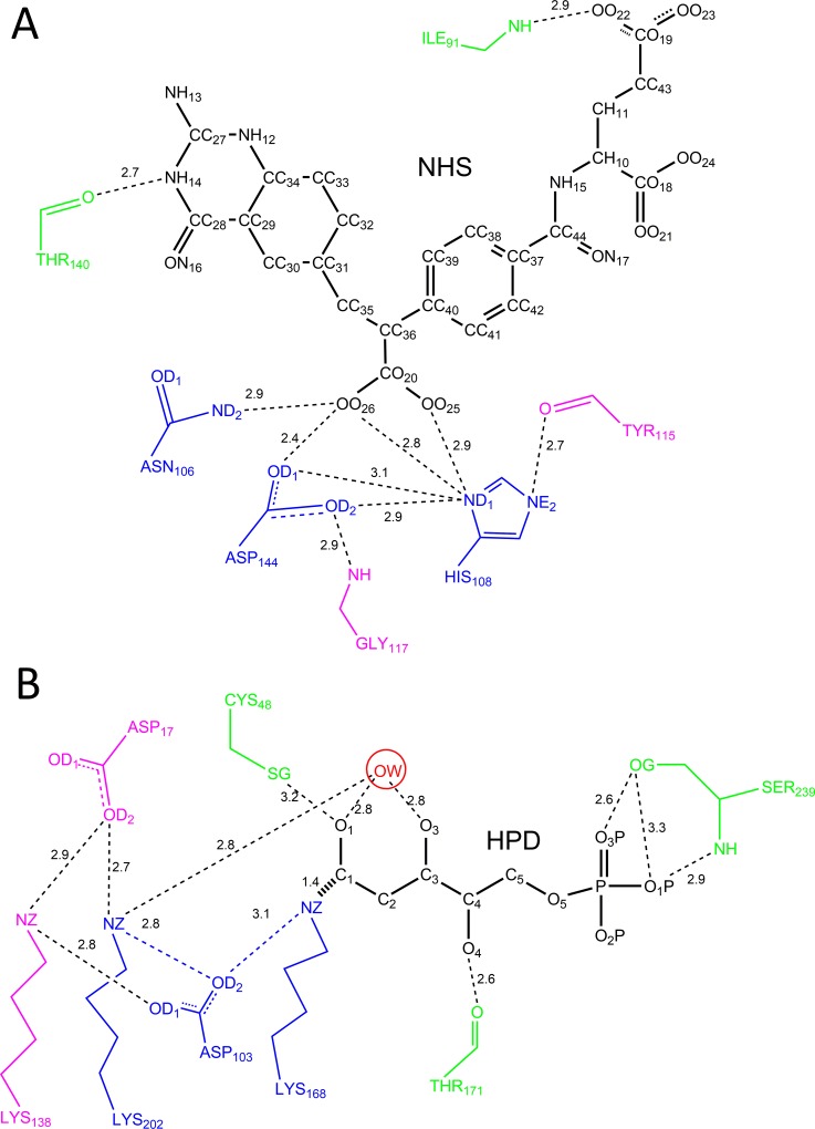
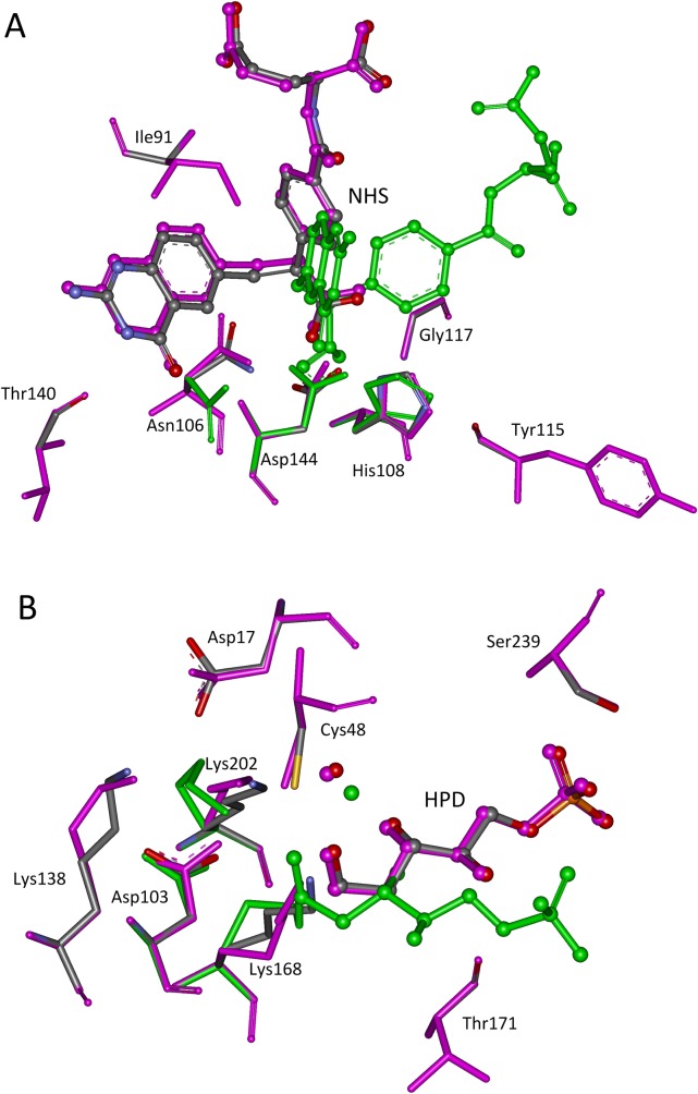
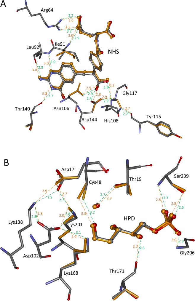
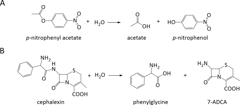
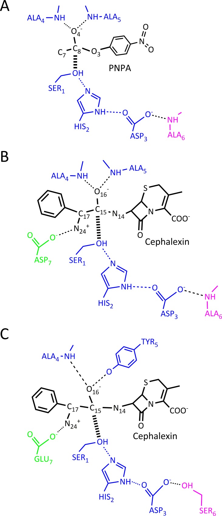
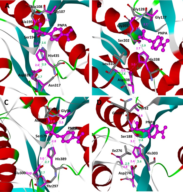
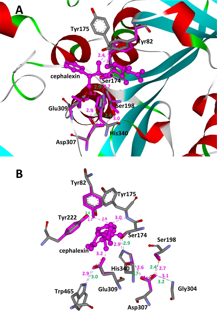
Active site preorganization helps native enzymes electrostatically stabilize the transition state better than the ground state for their primary substrates and achieve significant rate enhancement. In this report, we hypothesize that a complex active site model for active site preorganization modeling should help to create preorganized active site design and afford higher starting activities towards target reactions. Our matching algorithm ProdaMatch was improved by invoking effective pruning strategies and the native active sites for ten scaffolds in a benchmark test set were reproduced. The root-mean squared deviations between the matched transition states and those in the crystal structures were < 1.0 Å for the ten scaffolds, and the repacking calculation results showed that 91% of the hydrogen bonds within the active sites are recovered, indicating that the active sites can be preorganized based on the predicted positions of transition states. The application of the complex active site model for de novo enzyme design was evaluated by scaffold selection using a classic catalytic triad motif for the hydrolysis of p-nitrophenyl acetate. Eighty scaffolds were identified from a scaffold library with 1,491 proteins and four scaffolds were native esterase. Furthermore, enzyme design for complicated substrates was investigated for the hydrolysis of cephalexin using scaffold selection based on two different catalytic motifs. Only three scaffolds were identified from the scaffold library by virtue of the classic catalytic triad-based motif. In contrast, 40 scaffolds were identified using a more flexible, but still preorganized catalytic motif, where one scaffold corresponded to the α-amino acid ester hydrolase that catalyzes the hydrolysis and synthesis of cephalexin. Thus, the complex active site modeling approach for de novo enzyme design with the aid of the improved ProdaMatch program is a promising approach for the creation of active sites with high catalytic efficiencies towards target reactions.


Conflict of interest statement

Competing Interests: The authors have declared that no competing interests exist.

Figures

Fig 1
Fig 1
Schematic diagram of complex active site models for scaffolds: (A) 1c2t; (B) 1jcl. The catalytic residues are colored in blue, the residues that stabilize the catalytic residues are colored in pink, and the residues that stabilize the TS are colored in green.
Fig 2
Fig 2. Superposition of native and predicted active sites.
(A) 1c2t; (B) 1jcl. The transition states are shown in ball and stick model, and the active site residues in stick model. Atoms O, N, and C in crystal structures are colored in red, teal, and gray, respectively. The matched structures based on complex active site model are shown in pink, and in green for matched structures based on minimal active site model.
Fig 3
Fig 3
Side chain repacking results for scaffolds: (A) 1c2t; (B) 1jcl. The transition states are shown in ball and stick model, and the active site residues in stick model. Atoms O, N, and C in crystal structures are colored in red, teal, and gray, respectively. The matched TS and repacked residues are colored in orange. The hydrogen bonds in crystal structures are shown in dotted green lines, and the predicted hydrogen bonds are shown in dotted orange lines. The distances between hydrogen bonding donors and acceptors are shown in Å and labeled besides the dotted lines.
Fig 4
Fig 4. Reaction schemes of hydrolysis of PNPA and cephalexin.
PNPA: p-nitrophenyl acetate; 7-ADCA: 7-amino desacetoxycephalosporanic acid.
Fig 5
Fig 5. Complex active site models for PNPA and cephalexin based on different catalytic motifs.
(A) Classic catalytic triad motif for hydrolysis of PNPA; (B) Classic catalytic triad motif for hydrolysis of cephalexin; (C) Flexible catalytic triad motif for hydrolysis of cephalexin.
Fig 6
Fig 6. Superposition of native and predicted active sites for hydrolysis of PNPA on scaffolds.
(A) 1f6w; (B) 1jkm; (C) 1qe3; (D) 3m83. The transition states are shown in ball and stick model and colored in pink. The active site residues are shown in stick model. Atoms O, N, and C in crystal structures are colored in red, teal, and gray, respectively. Matched residues are colored in red. The hydrogen bonds in crystal structures are shown in dotted green lines, and the predicted hydrogen bonds are shown in dotted pink lines. The distances between hydrogen bonding donors and acceptors are shown in Å and labeled besides the dotted lines.
Fig 7
Fig 7. Matching results and active site residue sidechain repacking results in scaffold 1mpx.
(A) Superposition of native and matched active sites for hydrolysis of cephalexin on scaffold 1mpx. (B) Conformations of repacked residues based on matched cephalexin on scaffold 1mpx. The transition states are shown in ball and stick model and colored in pink. The active site residues are shown in stick model. Atoms O, N, and C in crystal structures are colored in red, teal, and gray, respectively. Matched residues are colored in red. The hydrogen bonds in crystal structures are shown in dotted green lines, and the predicted hydrogen bonds are shown in dotted pink lines. The distances between hydrogen bonding donors and acceptors are shown in Å and labeled besides the dotted lines.

References

    1. Frushicheva MP, Mills MJ, Schopf P, Singh MK, Prasad RB, Warshel A. Computer aided enzyme design and catalytic concepts. Curr Opin Chem Biol. 2014;21:56–62. 10.1016/j.cbpa.2014.03.022 - DOI - PMC - PubMed
    1. Schmid A, Dordick J, Hauer B, Kiener A, Wubbolts M, Witholt B. Industrial biocatalysis today and tomorrow. Nature. 2001;409(6817):258–68. - PubMed
    1. Bornscheuer U, Huisman G, Kazlauskas R, Lutz S, Moore J, Robins K. Engineering the third wave of biocatalysis. Nature. 2012;485(7397):185–94. 10.1038/nature11117 - DOI - PubMed
    1. Arnold FH. Design by directed evolution. Accounts of Chemical Research. 1998;31(3):125–31.
    1. Romero PA, Arnold FH. Exploring protein fitness landscapes by directed evolution. Nature Reviews Molecular Cell Biology. 2009;10(12):866–76. 10.1038/nrm2805 - DOI - PMC - PubMed

LinkOut - more resources