. 2016 May 31;11(5):e0156604.
doi: 10.1371/journal.pone.0156604. eCollection 2016.

Intrahepatic mRNA Expression of FAS, FASL, and FOXP3 Genes Is Associated with the Pathophysiology of Chronic HCV Infection

Affiliations

Intrahepatic mRNA Expression of FAS, FASL, and FOXP3 Genes Is Associated with the Pathophysiology of Chronic HCV Infection

Ednelza da Silva Graça Amoras et al. PLoS One. .

Abstract

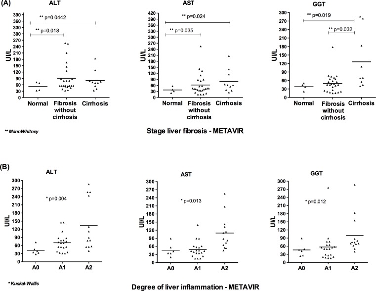
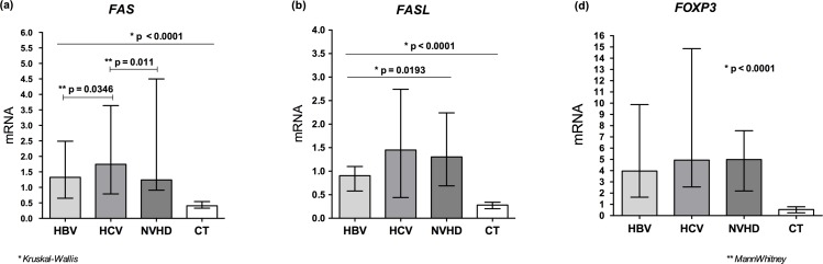
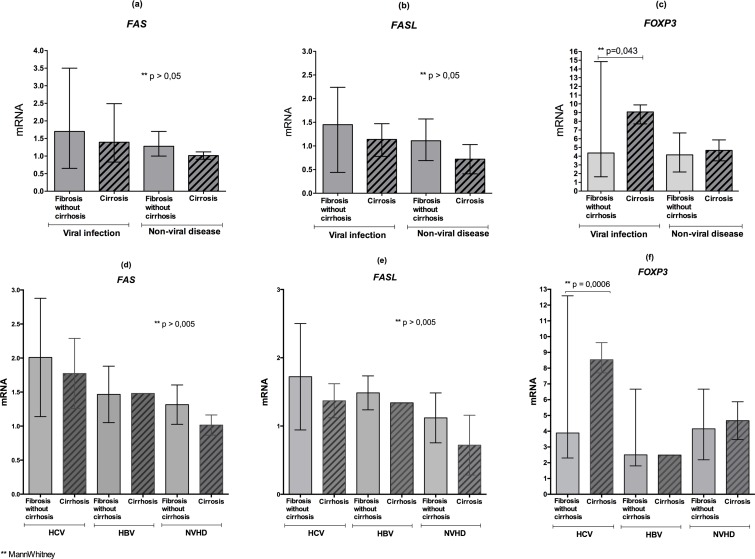
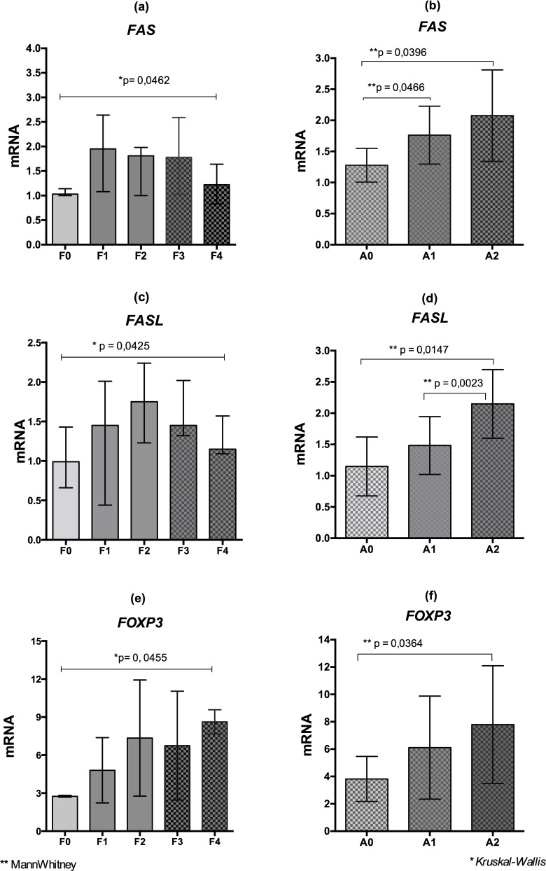
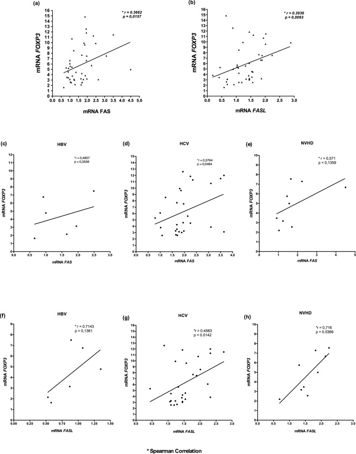
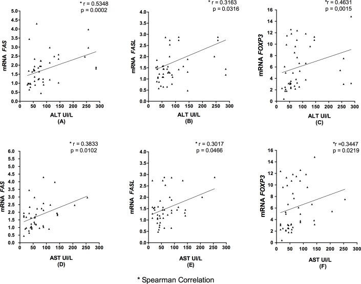
This study aimed to evaluate the relative mRNA expression of Fas receptor (FAS), Fas ligand (FASL), and forkhead box protein 3 (FOXP3) in liver biopsy specimens obtained from patients with viral and non-viral chronic hepatitis and correlate their expression with the fibrosis stage. A total of 51 liver biopsy specimens obtained from HBV (n = 6), HCV (n = 28), and non-viral hepatic disease (NVHD) (n = 9) patients and from individuals with normal liver histology (n = 8) (control-CT) were analyzed. Quantifications of the target genes were assessed using qPCR, and liver biopsies according to the METAVIR classification. The mRNA expression levels of FAS and FASL were lower in the CT group compared to the groups of patients. The increase in the mRNA expression of FAS and FASL was correlated with higher levels of inflammation and disease progression, followed by a decline in tissues with cirrhosis, and it was also associated with increased levels of alanine aminotransferase (ALT) and aspartate aminotransferase (AST). Higher mRNA expression of FOXP3 was observed in the HCV and NVHD groups, with the peak observed among patients with cirrhosis. The increased FOXP3 mRNA expression was positively correlated with increased FAS and FASL mRNA expression and the AST and ALT levels in all patients.

Conclusions: These results suggest that regardless of the cause, the course of chronic liver disease may be modulated by the analyzed genes and correlated with an increase in regulatory T cells during the liver damage followed by hepatocyte destruction by Fas/FasL system and subsequent non specific lymphocytic infiltrate accumulation.


Conflict of interest statement

Competing Interests: The authors have declared that no competing interests exist.

Figures

Fig 1
Fig 1. Mean plasma ALT, AST and GGT levels of all patients with viral and non-viral chronic hepatic disease.
A: patients without histological changes in the liver, with fibrosis (without cirrhosis) and with cirrhosis. B: according to the inflammatory activity.
Fig 2
Fig 2. Relative quantification mRNA levels FAS, FASL and FOXP3 in liver tissue.
A: mRNA levels FAS receptor in the groups of patients with HBV, HCV, and NVHD and the control group. B: mRNA levels FASL ligand in the groups of patients with HBV, HCV, and NVHD and the control group. C: mRNA levels FOXP3 in the groups of patients with HBV, HCV, and NVHD and the control group.
Fig 3
Fig 3. mRNA levels of FAS, FASL and FOXP3 in the groups without cirrhosis and with cirrhosis.
A-C: Quantification of the FAS receptor, FASL ligand and FOXP3 mRNA levels in the groups with fibrosis (without cirrhosis) and the groups with cirrhosis with viral and non-viral liver disease. D-F: Quantification of the FAS receptor, FASL ligand and FOXP3 mRNA levels in the groups with hepatic fibrosis (without cirrhosis) and cirrhosis due to viral and non-viral causes.
Fig 4
Fig 4. mRNA levels of FAS, FASL and FOXP3 according to the clinical conditions of liver.
A-F: mRNA levels of FAS, FASL and FOXP3 according to the fibrosis stages (F0 to F4) and inflammatory activity (A0 to A2) in the liver tissue of patients with viral and non-viral chronic liver disease (METAVIR).
Fig 5
Fig 5. mRNA levels of FAS, FASL and FOXP3 according to the clinical conditions of liver in the HCV infection.
A-F: mRNA levels of FAS, FASL and FOXP3 according to the fibrosis stages (F0 to F4) and inflammatory activity (A0 to A2) in the liver tissue of patients with HCV (METAVIR).
Fig 6
Fig 6. Correlation of FAS and FASL with FOXP3 mRNA levels in liver tissue.
A-E: Spearman correlation analysis with mRNA levels between FOXP3 and the FAS receptor and between FOXP3 and the FASL ligand in the chronic liver disease group (A and B), and according to the groups HBV (C and F), HCV infections (D and G), and NVHD (E and H).
Fig 7
Fig 7. Correlation of FAS, FASL and FOXP3 mRNA levels with ALT and AST concentrations.
A, D: Spearman correlation analysis between the FAS receptor mRNA levels and plasma ALT and AST concentrations. B, E: Spearman correlation analysis between the FASL mRNA levels and plasma ALT and AST concentrations. C, F: Spearman correlation analysis between the FOXP3 mRNA levels and plasma ALT and AST concentrations, in the chronic liver disease group.

References

    1. Guicciardi ME, Gores GJ. Apoptosis: a mechanism of acute and chronic liver injury. Gut 2005;54: 1024–1033. - PMC - PubMed
    1. Alter MJ Epidemiology of hepatitis C virus infection. World J Gastroenterol. 2007;13: 2436–2441. - PMC - PubMed
    1. Guidotti LG, Chisari FV. Immunobiology and pathogenesis of Viral hepatitis. Annu Rev Pathol. 2006;1: 23–61. - PubMed
    1. Mita E, Hayashi N, Lio S. Role of Fas ligand in apoptosis induced by hepatitis C virus infection. Biochem Biophys Res Commun. 1994;204: 468–474. - PubMed
    1. Jonuleit H, Schmitt E. The regulatory T cell family: distinct subsets and their interrelations. J Immunol. 2003;171: 6323–6327. - PubMed

MeSH terms