. 2016 May 31;11(5):e0156686.
doi: 10.1371/journal.pone.0156686. eCollection 2016.

HMGB1 Induces Secretion of Matrix Vesicles by Macrophages to Enhance Ectopic Mineralization

Affiliations

HMGB1 Induces Secretion of Matrix Vesicles by Macrophages to Enhance Ectopic Mineralization

Qiang Chen et al. PLoS One. .

Abstract

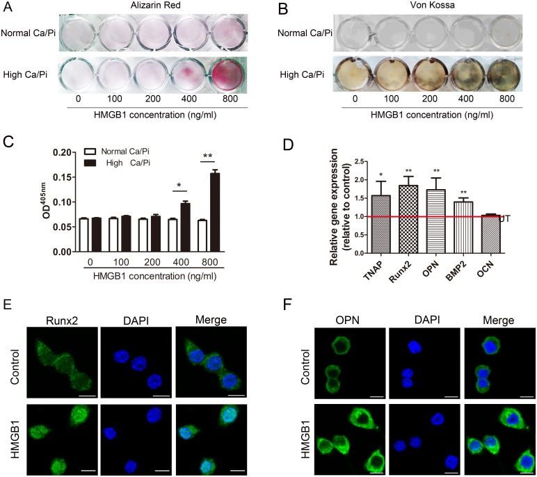
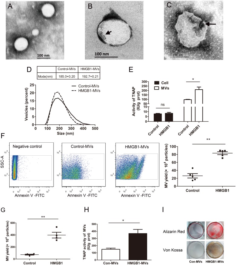
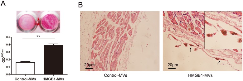
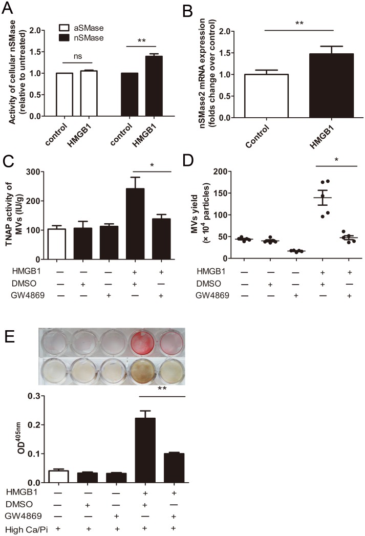
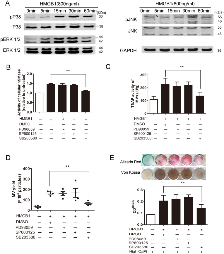
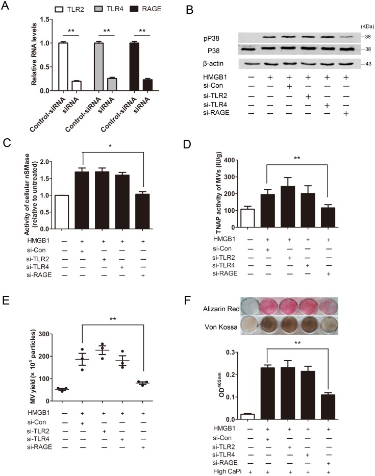
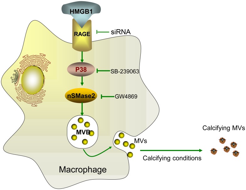
Numerous clinical conditions have been linked to ectopic mineralization (EM). This process of pathological biomineralization is complex and not fully elucidated, but thought to be started within matrix vesicles (MVs). We hypothesized that high mobility group box 1 (HMGB1), a cytokine associated with biomineralizing process under physiological and pathological conditions, induces EM via promoting MVs secretion from macrophages. In this study, we found that HMGB1 significantly promoted secretion of MVs from macrophages and subsequently led to mineral deposition in elevated Ca/Pi medium in vitro. Transmission electron microscopy of calcifying MVs showed formation of hydroxyapatite crystals in the vesicle interior. Subcutaneous injection into mice with MVs derived from HMGB1-treated cells showed a greater potential to initiate regional mineralization. Mechanistic experiments revealed that HMGB1 activated neutral sphingomyelinase2 (nSMase2) that involved the receptor for advanced glycation end products (RAGE) and p38 MAPK (upstream of nSMase2). Inhibition of nSMase2 with GW4869 or p38 MAPK with SB-239063 prevented MVs secretion and mineral deposition. Collectively, HMGB1 induces MVs secretion from macrophages at least in part, via the RAGE/p38 MAPK/nSMase2 signaling pathway. Our findings thus reveal a novel mechanism by which HMGB1 induces ectopic mineralization.


Conflict of interest statement

Competing Interests: The authors have declared that no competing interests exist.

Figures

Fig 1
Fig 1. HMGB1 promotes matrix mineralization of RAW264.7 cells.
Calcium deposit was visualized with Alizarin Red staining(A) and von Kossa staining (B), and Alizarin Red stains were eluted and measured at 405 nm (C). *P <0.05; **P <0.01, versus normal CaPi groups (n = 3). (D) Effects of HMGB1 on the mRNA expression of the mineralization-related markers TNAP, Runx2, OPN, BMP2 and OCN. The results were expressed as fold change of the untreated (UT) groups. (E and F) Effects of HMGB1 on the protein expression of the Runx2 and OPN detected by immunofluorescence staining. (Bar = 10 μm).
Fig 2
Fig 2. HMGB1 induces matrix vesicles secretion from RAW264.7 macrophages (A-F) or mouse peritoneal macrophages (G-I).
Transmission electron microscopic images show MVs with no crystalline hydroxyapatite (A), with early mineralized electron dense materials (short arrow) near the inner leaflet (B) and with matured crystal-like structures (long arrow) in the vesicle interior (C). (D) Size distribution of MVs was determined by dynamic light scattering method. (E) Assay of TNAP activity in cells (filled bar) or MVs (white bar). *P <0.05 (n = 3). (F) MVs secretion from cells was determined by flow cytometry. **P <0.01 (n = 5). (G) Effect of HMGB1 on secretion of MVs from mouse peritoneal macrophages was determined by flow cytometry. **P <0.01 (n = 4). (H) TNAP activity in MVs was determined. *P<0.05 (n = 4). (I) HMGB1-MVs from mouse peritoneal macrophages initiated mineralization in type I collagen-coated dishes as determined by Alizarin Red staining and von Kossa staining. For all grouped data, values are presented as the mean±SEM.
Fig 3
Fig 3. HMGB1-MVs initiate mineralization both in vitro and in vivo.
(A) Control-MVs or HMGB1-MVs from RAW264.7 cells initiated mineralization in type I collagen-coated dishes was determined by Alizarin Red staining and stains were eluted and measured at 405 nm. **P <0.01 (n = 3). (B) Hematoxylin and eosin (HE) staining of subcutaneous tissue sections 7 days after MVs injection. HMGB1-MVs induced calcium deposits (arrows) were associated with dermal collagen fibers. The presence of calcium deposits was confirmed using von Kossa stain. **P <0.01 (n = 5). For all grouped data, values are presented as the mean±SEM.
Fig 4
Fig 4. Blocking neutral sphingomyelinase-2 (nSMase) inhibited secretion of matrix vesicles and matrix mineralization induced by HMGB1.
(A) Cells were treated with 800 ng/ml HMGB1 for 24 h. The activity of nSMase and acid sphingomyelinase (aSMase) of cell homogenate were examined using a sphingomyelinase assay kit. The results are presented as a relative to control group after normalization by total protein content. **P <0.01 (n = 3). (B) Levels of nSMase2 mRNA expression were determined by real-time PCR. **P <0.01 (n = 3). (CE) Effects of inhibition of nSMase2 with GW4869 (5 μM) on HMGB1-induced MVs secretion and mineralization. TNAP activity in MVs (C) and MVs secretion (D) was measured. *P <0.05 (n = 5). (E) Mineralization was assessed by Alizarin Red staining and von Kossa staining. Alizarin Red stains were eluted and measured at 405 nm. DMSO (0.1%) was used as vehicle control. Values shown are the mean±SEM. **P <0.01 (n = 3).
Fig 5
Fig 5. Effects of MAP kinases signaling on HMGB1-induced nSMase2 activation, matrix vesicles releasing and mineralization.
(A) Cells were treated with 800 ng/ml HMGB1 for 0–60 min. The phosphorylated (upper row) and total (lower row) forms of ERK1/2, p38MAPK and JNK were assessed by western blot analysis. (B) Effects of inhibitors for ERK1/2 (PD98059), p38MAPK (SB203580) or JNK (SP600125) on the activity of nSMase. **P <0.01 (n = 3). (C and D) MVs were isolated from the culture supernatants, TNAP activity in MVs (C) and MVs secretion (D) were determined. **P <0.01 (n = 4). (E) Mineralization was assessed by Alizarin Red staining and von Kossa staining. Alizarin Red stains were eluted and measured at 405 nm. DMSO (0.1%) was used as vehicle control. Values shown are the mean±SEM **P <0.01 (n = 3).
Fig 6
Fig 6. HMGB1 signals through RAGE to promote p38MAPK/nSMase2 mediated secretion of matrix vesicles and mineralization.
(A) Real-time PCR analysis of mRNA expression in RAW264.7 cells after knockdown of TLR2, TLR4 or RAGE, respectively. **P <0.01 (n = 3). (B) Western blot analysis was performed for the p38MAPK phosphorylated forms and corresponding non-phosphorylated proteins. (C) nSMase activity of cells were detected at 24 h. *P <0.05 (n = 3). (D and E) MVs were isolated from the culture supernatants. TNAP activity in MVs (D) and MVs secretion (E) were determined. **P <0.01 (n = 3). (F) Mineralization was assessed by Alizarin Red staining and von Kossa staining, Alizarin Red stains were eluted and measured at 405 nm. Values shown are the mean±SEM **P <0.01 (n = 3).
Fig 7
Fig 7. Proposed role of HMGB1 in the secretion of matrix vesicles (MVs) by macrophages and subsequent mineralization.
MVs secretion by macrophages can be induced by HMGB1 through activation of neutral sphingomyelinase-2 (nSMase2) by the receptor for advanced glycation end products (RAGE)/p38 mitogen-activated protein kinase (p38MAPK) pathway. Pharmacological or genetic inhibition of RAGE, p38MAPK or nSMase2 is able to impede secretion of MVs. After being released into the extracellular space, MVs shift towards a pro-calcific state in a calcifying-prone condition.

References

    1. De Vilder EY, Vanakker OM. From variome to phenome: Pathogenesis, diagnosis and management of ectopic mineralization disorders. World J Clin Cases. 2015; 7:556–574. - PMC - PubMed
    1. Li Q, Jiang Q, Uitto J. Ectopic mineralization disorders of the extracellular matrix of connective tissue: Molecular genetics and pathomechanisms of aberrant calcification. Matrix Biol. 2014; 33:23–28. 10.1016/j.matbio.2013.06.003 - DOI - PMC - PubMed
    1. Avogaro A, Rattazzi M, Fadini GP. Ectopic calcification in diabetic vascular disease. Expert Opin Ther Targets. 2014; 18:595–609. 10.1517/14728222.2014.894021 - DOI - PubMed
    1. Otsuka F, Sakakura K, Yahagi K, Joner M, Virmani R. Has our understanding of calcification in human coronary atherosclerosis progressed? Arterioscler Thromb Vasc Biol. 2014; 34:724–736. 10.1161/ATVBAHA.113.302642 - DOI - PMC - PubMed
    1. Shapiro IM, Landis WJ, Risbud MV. Matrix vesicles: Are they anchored exosomes? Bone. 2015; 79:29–36. 10.1016/j.bone.2015.05.013 - DOI - PMC - PubMed

MeSH terms