. 2016 Jul-Aug;39(6):233-40.
doi: 10.1097/CJI.0000000000000128.

The Distribution of Human Stem Cell-like Memory T Cell in Lung Cancer

Affiliations

The Distribution of Human Stem Cell-like Memory T Cell in Lung Cancer

Hai Hong et al. J Immunother. 2016 Jul-Aug.

Abstract

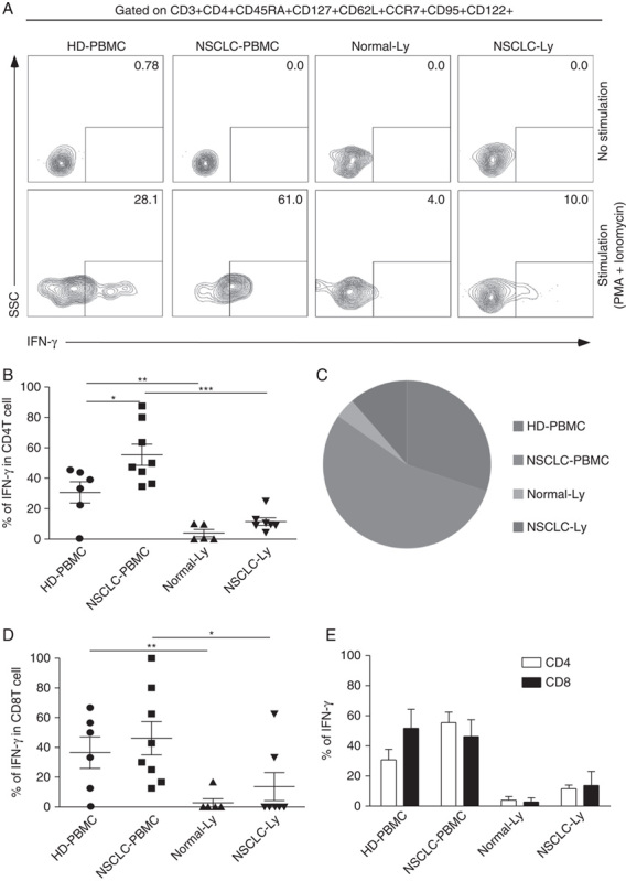
Human stem cell-like memory T (Tscm) cells are long-lived, self-renewing memory lymphocytes that can differentiate into effector cells and mediate strong antitumour response in murine model. The distribution and function of Tscm cells in human lung cancer remain unknown. In this study, we investigated the properties of human Tscm cells in the blood and lymph node of non-small cell lung cancer (NSCLC) patients. There were more CD4 Tscm cells in blood from NSCLC patients than from healthy donors, fewer CD4 and CD8 TSCM cells in blood than in lymph node from NSCLC patients. To further analyze their properties, we stimulated peripheral blood mononuclear cells from NSCLC patients by mitogens to examine cytokine production. Our data suggest that both CD4 and CD8 Tscm cells in blood produced interferon-γ significantly increased in NSCLC patients compare with healthy subjects. In addition, fewer Tscm cells produced interferon-γ in lymph node than in blood from NSCLC patients. Our results strongly suggest that the distribution and function of CD4 Tscm cells in NSCLC patients is upregulated. Understanding of the properties of stem-like memory T cells will supply a good rationale for designing the new adoptive immunotherapy in cancer.


Figures

FIGURE 1
FIGURE 1
Identification of Tscm CD4+CD45RA+CD45ROCD62L+CCR7+CD127+CD27+CD28+CD95+CD122+ T (Tscm) cell in human blood and lymph nodes. PBMCs were isolated from the blood of non–small cell lung cancer (NSCLC) patients (n=15) (NSCLC-PBMC) and healthy donors (n=11) (HD-PBMC); lymphocytes were isolated from the tumor-infiltrated lymph node of NSCLC patients who were collected blood at same time (n=7) (NSCLC-Ly); lymphocytes were isolated from the healthy lymph node of non lung cancer patients (n=7) (Normal-Ly), analyzed by flow cytometry. A, Representative flow cytometric analyses of CD4+CD45RA+CD45ROCD62L+CCR7+CD127+CD27+CD28+CD95+CD122+ T cells, indicating Tscm cells. B, The frequency of the CD4+Tscm cells in the HD-PBMC, NSCLC-PBMC, Normal-Ly, NSCLC-Ly. The events of CD4+Tscm cells in the blood and lymph node from NSCLC patients and healthy donors, expressed as the mean±SEM. C, The frequency of the CD8+Tscm cells in the HD-PBMC, NSCLC-PBMC, Normal-Ly, NSCLC-Ly, expressed as the mean±SEM. D, The events of Tscm of CD4+ and CD8+ cells in the blood and the lymph node from NSCLC patients and healthy donors. HD indicates healthy donors; IFN, interferon; PBMC, peripheral blood mononuclear cells; Tscm cell, stem cell–like memory T cell. *P<0.05; **P<0.005; ***P<0.001; Mann-Whitney test (2-tailed) and Student t test.
FIGURE 2
FIGURE 2
Cytokine production of the Tscm cell. HD-PBMCs (n=6), non–small cell lung cancer (NSCLC)-PBMCs (n=8) from the blood or lymphocytes isolated from Normal-Ly (n=5), NSCLC-Ly (n=7) were stimulated for 4–6 hours with PMA+ionomycin, and cytokine production was assessed by intracellular cytokine staining. A, Representative intracellular cytokine staining analysis of IFN-γ production by CD4+Tscm and CD8+Tscm cells in the blood and the lymph node relative to the unstimulated controls. B, The mean IFN-γ production (±SEM) of the CD4+ Tscm cells in the blood and lymph node from the nNSCLC patients and healthy donors. C, Pie charts depicting the quality of the IFN-γ-expressing CD4+Tscm cells in the blood and the lymph node from NSCLC patients and healthy donor. D, The mean IFN-γ production (±SEM) of the CD8+Tscm cells in the blood and lymph node from the NSCLC patients and healthy donors. E, The graph shows the relative frequencies of the IFN-γ-producing CD4+Tscm and CD8+Tscm cells in the HD-PBMC, LC-PBMC, Normal-Ly, NSCLC-Ly. HD indicates healthy donors; IFN, interferon; PBMC, peripheral blood mononuclear cells; PMA, phorbol 12-myristate13-acetate; Tscm cell, stem cell–like memory T cell; SSC, side scatter. *P<0.05; **P<0.005; ***P<0.001; Mann-Whitney test (2-tailed) and Student t test.
FIGURE 3
FIGURE 3
CD4+CD45RA+CD45ROCD95CD122+CD127+T cell population displays different phenotypes in human blood and lymph nodes. A, Flow cytometry plots showing IFN-γ expression in the CD4+CD45RA+/CD45ROCD95CD122+CD127+ T cells from the blood and lymph node of the non–small cell lung cancer (NSCLC) patients and healthy donors. B, The mean frequency (±SEM) of the events of CD4+CD45RA+/CD45ROCD95CD122+CD127+ T cells. C, The mean IFN-γ production (±SEM) of the CD4+CD45RA+/CD45ROCD95CD122+CD127+ T cells. HD indicates healthy donors; IFN, interferon; PBMC, peripheral blood mononuclear cells; PMA, phorbol 12-myristate13-acetate. *P<0.05; **P<0.005; paired Student t test.
FIGURE 4
FIGURE 4
CD4+CD45RA+CD95+ T cells and CD4+CD45RACD95+ T cells show distinct capacity for IFN-γ production. A, Representative analysis of IFN-γ production by CD4+CD45RA+CD95+ and CD4+CD45RACD95+T cells in the blood and the lymph node relative to the unstimulated controls from non–small cell lung cancer (NSCLC) patients and healthy donors. B, The mean IFN-γ production (±SEM) in the CD4+CD45RA+CD95+ T cells from the blood and lymph node of NSCLC patients and healthy donor. C, Representative analysis of IFN-γ production by CD4+CD45RACD95+ T cells in the blood and lymph node relative to the unstimulated controls from NSCLC patients and healthy donors. D, The percentage of IFN-γ-producing CD4+CD45RACD95+ T cells in the blood and lymph node from NSCLC patients and healthy donors, expressed as the mean±SEM. E, The plot shows the relative frequencies of the IFN-γ-producing CD4+CD45RA+CD95+ T cells and CD4+CD45RACD95+ T cells in the HD-PBMC, LC-PBMC, Normal-Ly, NSCLC-Ly. HD indicates healthy donors; IFN, interferon; PBMC, peripheral blood mononuclear cells; PMA, phorbol 12-myristate13-acetate; SSC, side scatter. *P<0.05; **P<0.005; ***P<0.001 Mann-Whitney test (2-tailed) and Student t test.

References

    1. Campbell JJ, Murphy KE, Kunkel EJ, et al. CCR7 expression and memory T cell diversity in humans. J Immunol. 2001;166:877–884. - PubMed
    1. Sallusto F, Lenig D, Forster R, et al. Two subsets of memory T lymphocytes with distinct homing potentials and effector functions. Nature. 1999;401:708–712. - PubMed
    1. Seder RA, Ahmed R. Similarities and differences in CD4+ and CD8+ effector and memory T cell generation. Nat Immunol. 2003;4:835–842. - PubMed
    1. Sathaliyawala T, Kubota M, Yudanin N, et al. Distribution and compartmentalization of human circulating and tissue-resident memory T cell subsets. Immunity. 2013;38:187–197. - PMC - PubMed
    1. Yu XZ, Anasetti C. Memory stem cells sustain disease. Nat Med. 2005;12:1282–1283. - PubMed

Publication types