. 2016 Jul 22;119(3):450-62.
doi: 10.1161/CIRCRESAHA.116.308870. Epub 2016 May 31.

TWIST1 Integrates Endothelial Responses to Flow in Vascular Dysfunction and Atherosclerosis

Affiliations

TWIST1 Integrates Endothelial Responses to Flow in Vascular Dysfunction and Atherosclerosis

Marwa M Mahmoud et al. Circ Res. .

Abstract

Rationale: Blood flow-induced shear stress controls endothelial cell (EC) physiology during atherosclerosis via transcriptional mechanisms that are incompletely understood. The mechanosensitive transcription factor TWIST is expressed during embryogenesis, but its role in EC responses to shear stress and focal atherosclerosis is unknown.

Objective: To investigate whether TWIST regulates endothelial responses to shear stress during vascular dysfunction and atherosclerosis and compare TWIST function in vascular development and disease.

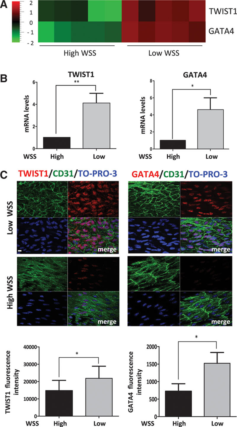
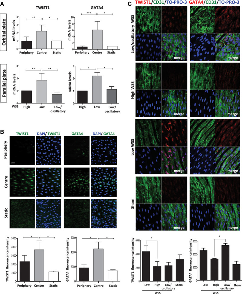
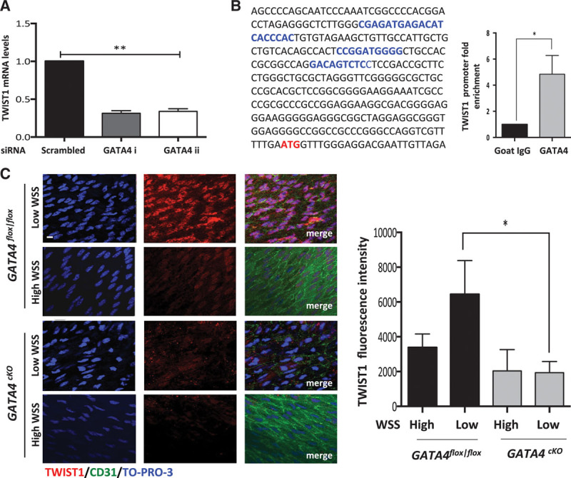
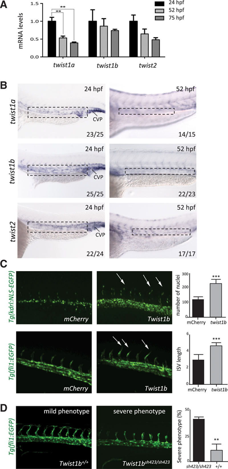
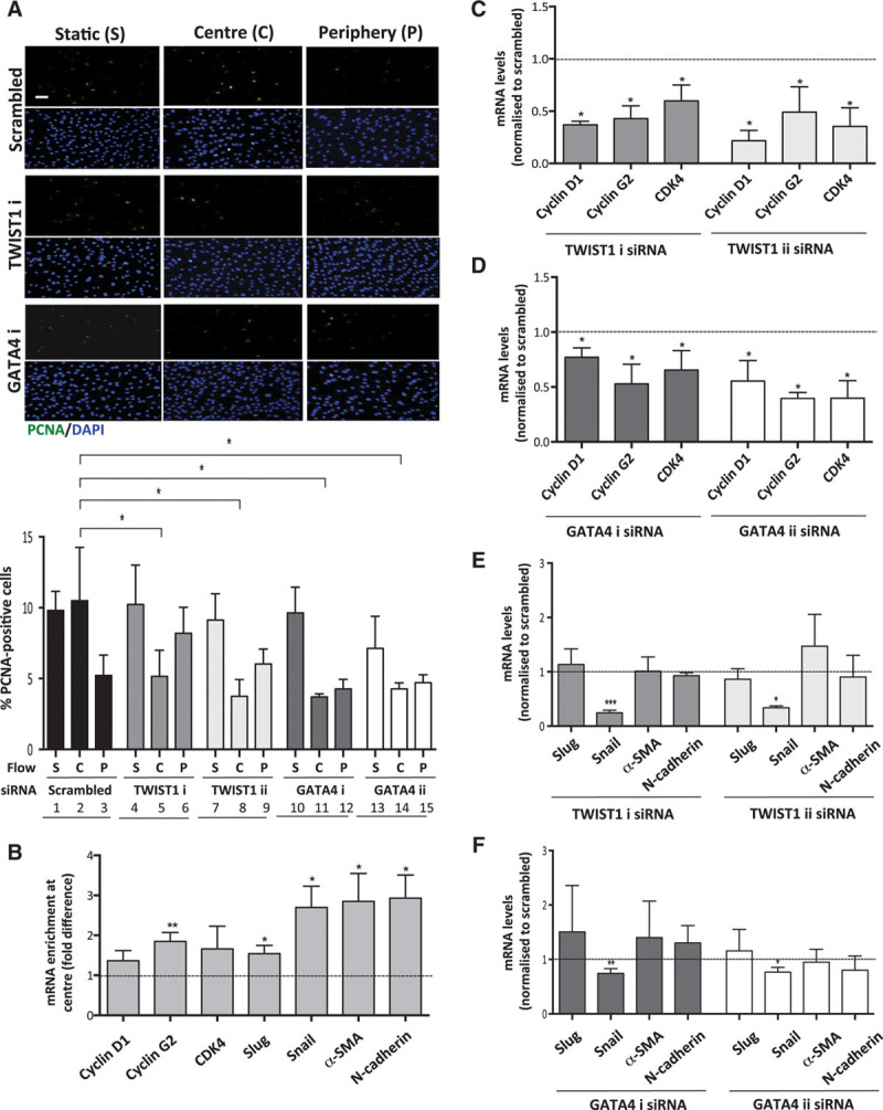
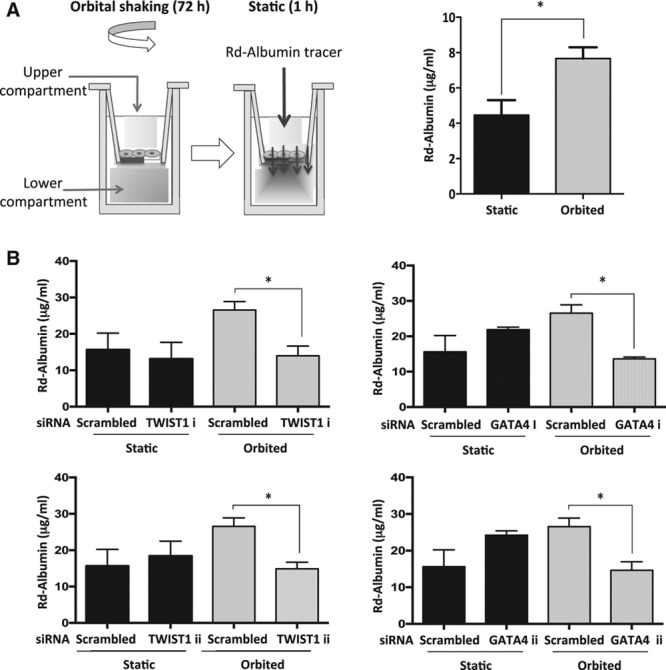
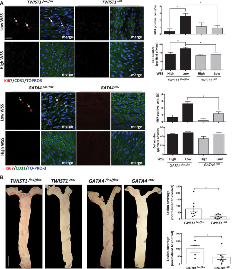
Methods and results: The expression and function of TWIST1 was studied in EC in both developing vasculature and during the initiation of atherosclerosis. In zebrafish, twist was expressed in early embryonic vasculature where it promoted angiogenesis by inducing EC proliferation and migration. In adult porcine and murine arteries, TWIST1 was expressed preferentially at low shear stress regions as evidenced by quantitative polymerase chain reaction and en face staining. Moreover, studies of experimental murine carotid arteries and cultured EC revealed that TWIST1 was induced by low shear stress via a GATA4-dependent transcriptional mechanism. Gene silencing in cultured EC and EC-specific genetic deletion in mice demonstrated that TWIST1 promoted atherosclerosis by inducing inflammation and enhancing EC proliferation associated with vascular leakiness.

Conclusions: TWIST expression promotes developmental angiogenesis by inducing EC proliferation and migration. In addition to its role in development, TWIST is expressed preferentially at low shear stress regions of adult arteries where it promotes atherosclerosis by inducing EC proliferation and inflammation. Thus, pleiotropic functions of TWIST control vascular disease and development.

Keywords: atherosclerosis; cholesterol; gastrulation; obesity; transcription factors.


Figures

Figure 1.
Figure 1.
TWIST1 and GATA4 were preferentially expressed at low shear atherosusceptible sites. A and B, Expression of TWIST1 and GATA4 was studied at low wall shear stress (WSS; inner curvature) and high WSS (outer curvature) regions of the porcine aorta. A, Differentially expressed genes were identified using microarrays (n=5 pigs) and the expression patterns of TWIST1 and GATA4 are presented as a heat map with red indicating enrichment in expression. B, Results were validated by qRT-PCR of a different cohort of pigs (n=5). The expression level at the low WSS site is presented relative to the expression at the high WSS site (normalized to 1; dotted line). Mean levels±SEM are shown. C, Expression levels of TWIST1 or GATA4 in EC were assessed by en face staining of low (inner curvature) or high (outer curvature) WSS regions of the aorta in C57BL/6 mice (red). Endothelial cells (ECs) were identified by costaining with anti-CD31 antibodies conjugated to FITC (fluorescein isothiocyanate; green). Cell nuclei were identified using TO-PRO-3 (blue). Representative images and quantification of TWIST1 or GATA4 (mean±SEM) are shown. *P<0.05 and **P<0.01, using a paired t test.
Figure 2.
Figure 2.
Low shear stress induced TWIST1 and GATA4. A, Human umbilical vein endothelial cells (HUVECs) were exposed to orbital flow to generate low (center) or high (periphery) wall shear stress (WSS) or were cultured under static conditions. Alternatively, HUVECs were exposed to high, low, or low/oscillatory WSS using a parallel-plate system. After 72 h, levels of TWIST1 or GATA4 transcripts were quantified by qRT-PCR. B, HUVECs were exposed to orbital flow to generate low (center) or high (periphery) WSS for 72 h or were cultured under static conditions. Expression of TWIST1 and GATA4 was determined by immunofluorescent staining (green) and costaining using DAPI (4′,6-diamidino-2-phenylindole; blue). Bar=50 μm. Fluorescence intensities were measured in multiple cells in 3 independent experiments, and mean values±SEM are shown. C, Flow-altering, constrictive cuffs were placed on the right carotid arteries of C57BL/6 mice. They generated anatomically distinct regions exposed to low, high, and low oscillatory WSS (as indicated). Right (experimental) and left (sham-operated) carotid arteries were harvested after 14 days, and en face staining was performed using anti-TWIST1 or anti-GATA4 antibodies (red), anti-CD31 antibodies conjugated to FITC (fluorescein isothiocyanate; green), and the nuclear counter stain TO-PRO-3 (blue). Representative images and quantification of TWIST1 or GATA4 expression (mean±SEM) are shown. Bar=10 μm. Data were pooled from 5–6 independent experiments. *P<0.05 and **P<0.01, using a 1-way ANOVA.
Figure 3.
Figure 3.
GATA4 induced TWIST1 in response to low shear stress. A, Human umbilical vein endothelial cells (HUVECs) were transfected with 2 different small interfering RNAs (siRNAs) targeting GATA4 or with scrambled sequences and exposed to orbital flow for 72 h. Cells exposed to low wall shear stress (WSS; center of well) were collected, and TWIST1 transcript levels were quantified by qRT-PCR. B, GATA4-binding sites (A/T)GATAA(G) were predicted in the TWIST1 5′ untranslated region, and their position (blue text) in relation to the translational start site (red text) is shown. HUVECs were exposed to orbital flow for 72 h before chromatin immunoprecipitation using anti-GATA4 or irrelevant isotype control antibodies. The levels of TWIST1 promoter DNA were assessed by qPCR and fold enrichment in anti-GATA4 compared with control precipitates was calculated. Mean levels±SEM are shown. A and B, Data from at least 3 independent experiments were pooled. *P<0.05 and **P<0.01 using an unpaired t test. C, Endothelial cells (ECs) at low WSS (susceptible) or high WSS (protected) regions of the aorta in GATA4cKO or GATA4fl/fl mice were studied by en face staining (n=5). TWIST1 (red) expression was assessed by en face staining. Representative images are shown, and TWIST1 expression was quantified (mean fluorescence±SEM is shown). Bar=10 μm. *P<0.05 using a 2-way ANOVA.
Figure 4.
Figure 4.
twist promoted intersegmental vessel sprouting in zebrafish embryos. A, The expression of twist1a, twist1b, and twist2 was studied at 24 to 75 hours post fertilization (hpf) by qPCR of the trunk and tail of embryos. Data were pooled from n≥15 embryos studied in 3 independent experiments and mean values±SEs are shown. B, The expression of twist1a, twist1b, and twist2 was studied at 24 and 52 hpf by in situ hybridization. Data are representative of the majority of embryos analyzed (proportion indicated lower right of each part) and were closely similar in at least 3 independent experiments. Higher magnification insets are shown (marked in top). Bar=100 µm. C, Zebrafish embryos (wild-type, Tg(fli1:EGFP), or Tg(kdrl:NLS-EGFP)) were treated with twist1b mRNA (to enforce expression) or treated with mCherry mRNA as a control. They were studied at 24 to 27 hpf using confocal microscopy to visualize endothelial cell (EC) nuclei (Tg(kdrl:NLS-EGFP); top) or angiogenic sprouts (Tg(fli1:EGFP); bottom; arrows). Representative images are shown. Cell numbers and the length of intersegmental vessels (ISVs; third to fifth vessels in the field view) were quantified in multiple embryos, and mean values±SEM are shown (right). D, The twist1b coding sequence was mutated by introduction of a 4 bp deletion causing a frameshift and premature stop (mutant allele designated twist1bsh423). twist1bsh423/+ Tg(fli1:EGFP) fish were incrossed, and embryos were treated with a morpholino directed against twist1a. Sprouting of ISV was assessed at 34 hpf. Embryos were classified into those that displayed minimal sprouting (severe phenotype) and those with intermediate levels of sprouting (mild phenotype). Genotyping was subsequently performed, and the proportion of twist1b homozygous mutants (twist1bsh423/sh423) and twist1b homozygous wild-types (twist1b+/+) in the severe phenotype group was calculated (% indicated). Representative images are shown. Data were closely similar in 3 independent experiments. ***P<0.001 and **P<0.01 using a 1-way ANOVA (A) or unpaired t test (C and D). CVP indicates caudal vein plexus.
Figure 5.
Figure 5.
TWIST1 and GATA4 promoted proliferation in endothelial cells (ECs) exposed to low shear stress. Human umbilical vein endothelial cells (HUVECs) were treated with 2 different small interfering RNAs (siRNAs) targeting TWIST1 or GATA4 (designated i and ii) or with scrambled nontargeting siRNA or remained untransfected. Cells were subsequently cultured in 6-well plates before exposure to orbital flow to generate low (center, C) or high (periphery, P) wall shear stress (WSS) for 72 h. Alternatively, cells were maintained under static (S) conditions. A, Cell proliferation was quantified by immunofluorescent staining using anti-proliferating cell nuclear antigen (PCNA) antibodies and costaining using DAPI (4′,6-diamidino-2-phenylindole). Images are representative of those generated in 3 independent experiments using 1 version of the gene-specific siRNA or scrambled control sequences (bar=50 μm). The percentage of PCNA-positive cells were calculated for multiple fields of view in at least 3 independent experiments, and mean values±SEM are shown. *P<0.05 using a 2-way ANOVA. B–F, The expression of cell cycle regulators and endothelial–mesenchymal transition genes was quantified using qRT-PCR. B, The expression level in cells at the center (low WSS) is presented relative to the expression at the periphery (high WSS; normalized to 1; dotted line). C–F, Transfected cells were exposed to low WSS (center). The expression level in cells transfected with gene-targeting siRNA is presented relative to the expression in cells transfected with scrambled control siRNA (normalized to 1; dotted line). Data were pooled from 3 independent experiments, and mean values±SEM are shown. *P<0.05, **P<0.01, and ***P<0.001 using an unpaired t test.
Figure 6.
Figure 6.
TWIST1 and GATA4 promoted permeability in endothelial cells (ECs) exposed to low shear stress. The influence of TWIST1 and GATA4 on EC permeability under low wall shear stress (WSS) was studied. A, Cells cultured on transwell inserts were exposed to orbital flow (low WSS) or static conditions for 72 h before assessment of endothelial permeability under static conditions using rhodamine (Rd) albumin as a tracer. A schematic is shown (left). The concentration of Rd-albumin in the lower compartment was measured, and mean values±SEM are shown (right). B, Human umbilical vein endothelial cells (HUVECs) were treated with 2 different small interfering RNAs (siRNAs) targeting TWIST1 or GATA4 (designated i and ii) or with scrambled nontargeting siRNA. Transfected cells cultured on transwell inserts were exposed to orbital flow (low WSS) or static conditions for 72 h before assessment of endothelial permeability using Rd-albumin. The concentration of Rd-albumin in the lower compartment was measured in 3 independent experiments, and mean values±SEM are shown. *P<0.05 using a paired t test.
Figure 7.
Figure 7.
TWIST1 and GATA4 promote endothelial cell (EC) proliferation and lesions at low shear stress sites. A, EC at low wall shear stress (WSS; susceptible) or high WSS (protected) regions of the aorta were studied by en face staining in TWIST1cKO or TWIST1flox/flox mice (top) or in GATA4cKO or GATA4flox/flox mice (bottom). EC proliferation was quantified by anti-Ki67 staining (red). EC were identified by costaining with anti-CD31 antibodies (green). Cell nuclei were identified using TOPRO3 (blue). The proportion of Ki67-positive cells, and number of cells was calculated and mean values±SEM are shown. Bar=10 μm. Data were pooled from 5 independent experiments. B, TWIST1cKO or TWIST1flox/flox mice (right) or GATA4cKO or GATA4flox/flox mice (left) were treated with AAV-PCSK9. After 1 wk, they were exposed to a Western diet for 6 wk. Lesions were stained using Oil Red O and quantified using image J software. Representative images are shown (Bar=1 mm). The percentage of lesion coverage was calculated. Data were pooled from multiple mice and mean values±SEM are shown. **P<0.01 and *P<0.05 using a 2-way ANOVA (A) or unpaired t test (B).

References

    1. Kwak BR, Bäck M, Bochaton-Piallat ML, et al. Biomechanical factors in atherosclerosis: mechanisms and clinical implications. Eur Heart J. 2014;35:3013–3020, 3020a. doi: 10.1093/eurheartj/ehu353. - PMC - PubMed
    1. Suo J, Ferrara DE, Sorescu D, Guldberg RE, Taylor WR, Giddens DP. Hemodynamic shear stresses in mouse aortas: implications for atherogenesis. Arterioscler Thromb Vasc Biol. 2007;27:346–351. doi: 10.1161/01.ATV.0000253492.45717.46. - PubMed
    1. Dai G, Kaazempur-Mofrad MR, Natarajan S, Zhang Y, Vaughn S, Blackman BR, Kamm RD, García-Cardeña G, Gimbrone MA., Jr Distinct endothelial phenotypes evoked by arterial waveforms derived from atherosclerosis-susceptible and -resistant regions of human vasculature. Proc Natl Acad Sci USA. 2004;101:14871–14876. doi: 10.1073/pnas.0406073101. - PMC - PubMed
    1. Passerini AG, Polacek DC, Shi C, Francesco NM, Manduchi E, Grant GR, Pritchard WF, Powell S, Chang GY, Stoeckert CJ, Jr, Davies PF. Coexisting proinflammatory and antioxidative endothelial transcription profiles in a disturbed flow region of the adult porcine aorta. Proc Natl Acad Sci USA. 2004;101:2482–2487. - PMC - PubMed
    1. Hajra L, Evans AI, Chen M, Hyduk SJ, Collins T, Cybulsky MI. The NF-kappa B signal transduction pathway in aortic endothelial cells is primed for activation in regions predisposed to atherosclerotic lesion formation. Proc Natl Acad Sci USA. 2000;97:9052–9057. - PMC - PubMed

Publication types

MeSH terms