Review
. 2016 Jul 15;56(7):416-41.
doi: 10.2176/nmc.ra.2016-0020. Epub 2016 May 27.

Research into the Physiology of Cerebrospinal Fluid Reaches a New Horizon: Intimate Exchange between Cerebrospinal Fluid and Interstitial Fluid May Contribute to Maintenance of Homeostasis in the Central Nervous System

Affiliations
Review

Research into the Physiology of Cerebrospinal Fluid Reaches a New Horizon: Intimate Exchange between Cerebrospinal Fluid and Interstitial Fluid May Contribute to Maintenance of Homeostasis in the Central Nervous System

Mitsunori Matsumae et al. Neurol Med Chir (Tokyo). .

Erratum in

Abstract

Cerebrospinal fluid (CSF) plays an essential role in maintaining the homeostasis of the central nervous system. The functions of CSF include: (1) buoyancy of the brain, spinal cord, and nerves; (2) volume adjustment in the cranial cavity; (3) nutrient transport; (4) protein or peptide transport; (5) brain volume regulation through osmoregulation; (6) buffering effect against external forces; (7) signal transduction; (8) drug transport; (9) immune system control; (10) elimination of metabolites and unnecessary substances; and finally (11) cooling of heat generated by neural activity. For CSF to fully mediate these functions, fluid-like movement in the ventricles and subarachnoid space is necessary. Furthermore, the relationship between the behaviors of CSF and interstitial fluid in the brain and spinal cord is important. In this review, we will present classical studies on CSF circulation from its discovery over 2,000 years ago, and will subsequently introduce functions that were recently discovered such as CSF production and absorption, water molecule movement in the interstitial space, exchange between interstitial fluid and CSF, and drainage of CSF and interstitial fluid into both the venous and the lymphatic systems. Finally, we will summarize future challenges in research. This review includes articles published up to February 2016.


Conflict of interest statement

Conflicts of Interest Disclosure

The authors declare no conflicts of interest.

Figures

Fig. 1
Fig. 1
a: Albrecht von Haller (1708–1777) is a Swiss anatomist and physiologist. His book, Primae lineae physiologiae in usum praelectionum academicarum. Gottingae: A. Vandenhoeck (1747), is shown. Von Haller, who was given credit for the discovery of cerebrospinal fluid (CSF) by Domenico Cotugno, stated interesting anatomical findings including the observation that the consistency of CSF increases after death (Public domain). b: Domenico Felice Antonio Cotugno (1736–1822) is a Neapolitan anatomist. His book, De ischiade nervosa commentarius. Viennae: Apud Rudolphum Gräffer (1770), is shown. Cotugno reported that liquid is present and air bubbles are absent at the meninges when it is incised and opened carefully. Therefore, Cotugno postulated that the presence of CSF at the spinal cord and brain surface may have been overlooked with the conventional cervical decapitation method. Furthermore, Cotugno also confirmed the outflow of liquid when a drain is placed in the lumbar sac of a cadaver in the standing position. A notable aspect of the work by Cotugno is that he proved the presence of CSF in both the cranial cavity and the spinal cavity (Public domain). c: Emanuel Swedenborg (1688–1772) is an anatomist with a degree in mining engineering. Swedenborg described CSF using terms such as “spirituous lymph” and “highly gifted juice.” His book, The Cerebrum and Its Parts. London: James Speirs (1882), is shown (Public domain).
Fig. 2
Fig. 2
The front cover ( left ) and part of page 8 ( right ) of “liquide céphalo-rachidien ou cérébro-spinal” described by François Jean Magendie owned by Kyoto University Library are shown. The foramen of Magendie that exists at the exit of the fourth ventricle is named after this scientist, who is well known for demonstrating the connection between the ventricular system and subarachnoid space. However, he is also famous for using the term “Le liquid cérébro-spinal” for the first time ( red underlined portion in right figure). Photos reprinted with the permission of Kyoto University Library.
Fig. 3
Fig. 3
The front cover of Key A, Retzius G. Studien in der anatomie des nervensystems und des bindegewebes. Stockholm: Norstedt & Söner (1875) owned by Niigata University Library is shown. This article is always referenced when cerebrospinal fluid (CSF) absorption from the arachnoid granulation or villi is described. The detailed illustrations of the cerebral superficial subarachnoid space and Virchow-Robin space are key characteristics of this article. There are illustrations in which CSF eliminated from the subarachnoid space is present in the retro-orbital tissue and cervical lymphatic system, and there are also descriptions of anatomical findings that are widely suggestive of CSF absorption. Images of the copies are taken with the permission of Niigata University Library.
Fig. 4
Fig. 4
Tafel XXIX Figure 4 ( upper panel ) and Tafel XXVIII Figure 2 ( lower panel ) of the first volume of “Studien in der anatomie des nervensystems und des bindegewebes. Stockholm: Norstedt & Söner (1875) by Key A, Retzius G. owned by Niigata University Library are shown. Detailed figures of the arachnoid villi that protrude from the subarachnoid space are depicted. Images of copies are taken with the permission of Niigata University Library.
Fig. 5
Fig. 5
Cerebrospinal fluid (CSF) exit pathways from the subarachnoid space are shown. The first pathway shows absorption by the arachnoid granulation or villi and subsequent exit through the venous sinus. The second pathway shows CSF migration from the meningeal lymphatic vessels to the cervical lymph node. There are other routes in which CSF reaches the cervical lymph node from the cribriform plate via the nasal mucosa.
Fig. 6
Fig. 6
A schematic diagram of cerebrospinal fluid (CSF) and interstitial fluid exchange among the ventricle, subarachnoid space reservoir, and brain parenchyma is shown. Glia, which covers the neurovascular unit, is located at the border of the area in which water enters and exits the brain and spinal cord, and water is exchanged at the aquaporin channel of astrocyte foot processes or at other sites through the endothelium via diffusion or vesicular transport. Water movement at the ependymal layer, pia mater, and Virchow-Robin space is bidirectional. The right cerebral hemisphere has a mixing of interstitial fluid secreted within the brain parenchyma and CSF that enters the brain parenchyma, and subsequent drainage from the brain parenchyma into the CSF reservoir (subarachnoid space and ventricles). The left cerebral hemisphere shows that CSF penetrates from the ventricles and subarachnoid space into the brain parenchyma.
Fig. 7
Fig. 7
Aquaporins (AQPs) are proteins with a molecular weight of 26–30 kDa and are composed of ∼250–290 amino acid residues. AQPs pass through the membrane six times in the vicinity of the pore (membrane-spanning) and allow water to travel in both directions through this pore. Reprinted with permission from Reference Figure 1 . (Zelenina M: Regulation of brain aquaporins. Neurochem Int 57: 468–488, 2010).
Fig. 8
Fig. 8
Some aquaporins (AQPs) contain a structure called the NPA (asparagine-proline-alanine) motif. The center portion of this motif has an hourglass-shaped structure, which penetrates through the lipid bilayer. Substance transport occurs here. AQP1 only allows water to pass through. At this hourglass-shaped portion, the diameter is narrowest at approximately 2.8 Å, which is the size in which one water molecule can just pass through. Water molecules can traverse in both directions at this site. Reprinted with the permission from Reference Figure 1 . (Badaut J, Lasbennes F, Magistretti PJ, Regli L: Aquaporins in brain: distribution, physiology, and pathophysiology. J Cereb Blood Flow Metab 22: 367–378, 2002).
Fig. 9
Fig. 9
An axial slice of the cerebrum from a cadaver shows that the distance from the subarachnoid space to the lateral ventricular wall as well as the distance from the Sylvian fissure to the third ventricular wall is almost equal in each region ( solid lines ). Cerebrospinal fluid (CSF) and interstitial fluid travel a long distance when the distal part of the Sylvian fissure is not present between the third ventricle and the convexity of the subarachnoid space ( dashed line ). Reprinted with the permission from Reference modified Figure 1 . [Sato O: [Reconsideration of research into cerebrospinal fluid], in Arai, H, Ishikawa, M, Mori, E ( eds ): iNPH: Idiopathic Normal Pressure Hydrocephalus . Kyoto, Kinpodo, 2014, pp 8–18 (Japanese)].

References

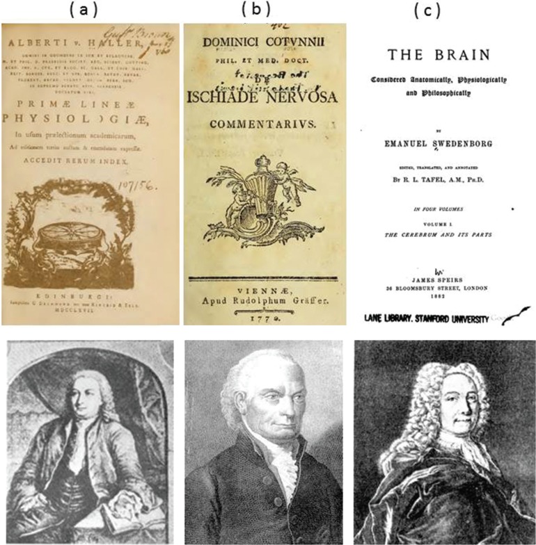
    1. Di leva A, Yaşargil MG: Liquor cotunnii: the history of cerebrospinal fluid in Domenico Cotugno’s work. Neurosurgery 63: 352– 358; discussion 358, 2008. - PubMed
    1. Deisenhammer F: The history of cerebrospinal fluid, in Deisenhammer F, Sellebjerg F, Teunissen CE, Tumani H. (eds): Cerebrospinal Fluid in Clinical Neurology. Cham, Springer International Publishing, 2015, pp 3–16
    1. Cotugno D: De ischiade nervosa commentarius. Viennae, Apud Rudolphum Gräffer, 1770.
    1. Swedenborg E: The Cerebrum and its Parts, Vol 1. London, Swedenborg Library Academy of the New Church, 1882.
    1. von Haller A: Primae lineae physiologiae in usum praelectionum academicarum. Göttinten, Vandenhoeck, 1747.

MeSH terms