. 2016 May 31:17:238.
doi: 10.1186/s12891-016-1094-8.

Coordinate and synergistic effects of extensive treadmill exercise and ovariectomy on articular cartilage degeneration

Affiliations

Coordinate and synergistic effects of extensive treadmill exercise and ovariectomy on articular cartilage degeneration

Kazumasa Miyatake et al. BMC Musculoskelet Disord. .

Abstract

Background: Although osteoarthritis (OA) is a multifactorial disease, little has been reported regarding the cooperative interaction among these factors on cartilage metabolism. Here we examined the synergistic effect of ovariectomy (OVX) and excessive mechanical stress (forced running) on articular cartilage homeostasis in a mouse model resembling a human postmenopausal condition.

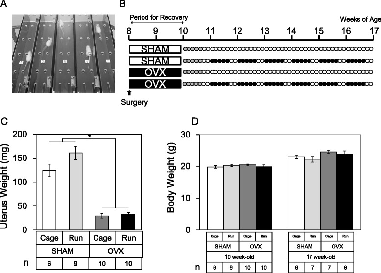
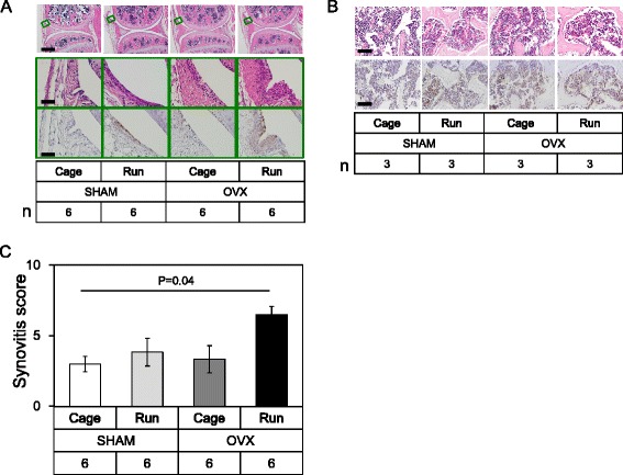
Methods: Mice were randomly divided into four groups, I: Sham, II: OVX, III: Sham and forced running (60 km in 6 weeks), and IV: OVX and forced running. Histological and immunohistochemical analyses were performed to evaluate the degeneration of articular cartilage and synovitis in the knee joint. Morphological changes of subchondral bone were analyzed by micro-CT.

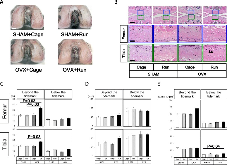
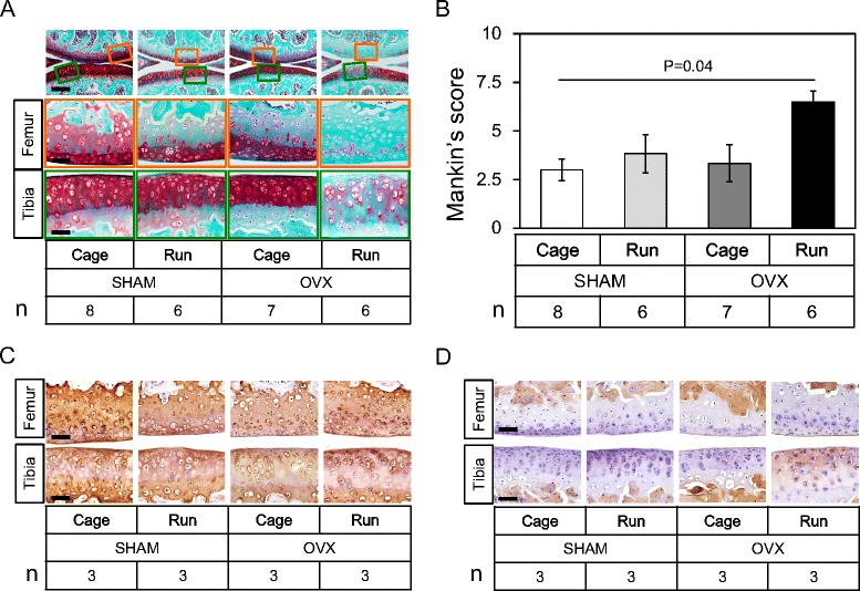
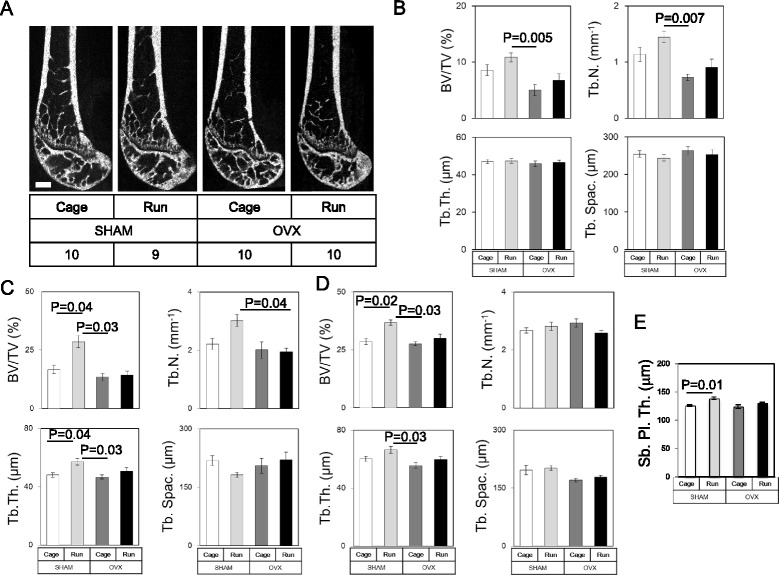
Results: Micro-CT analyses showed significant loss of metaphyseal trabecular bone volume/tissue volume (BV/TV) after OVX as described previously. Forced running increased the trabecular BV/TV in all mice. In the epiphyseal region, no visible alteration in bone morphology or osteophyte formation was observed in any of the four groups. Histological analysis revealed that OVX or forced running respectively had subtle effects on cartilage degeneration. However, the combination of OVX and forced running synergistically enhanced synovitis and articular cartilage degeneration. Although morphological changes in chondrocytes were observed during OA initiation, no signs of bone marrow edema were observed in any of the four experimental groups.

Conclusion: We report the coordinate and synergistic effects of extensive treadmill exercise and ovariectomy on articular cartilage degeneration. Since no surgical procedure was performed on the knee joint directly in this model, this model is useful in addressing the molecular pathogenesis of naturally occurring OA.

Keywords: Forced running; Joint inflammation; Mechanical stress; Naturally occurring osteoarthritis model; Ovariectomy.


Figures

Fig. 1
Fig. 1
Study design. a A picture of the treadmill apparatus used in this study. b Schematic overview of the experimental protocol. Female Balb/cCrSlc mice were randomly divided into four groups. At 8 weeks of age, mice underwent OVX or SHAM surgery with 2 weeks for recovery. At 10 weeks of age, mice were subjected to a training period of forced running at 12 m/s for 10 min as an adaptation to treadmill running (5 days a week, gray circle). After the initial training period, “Run” groups were then subjected to forced running at 12 m/s for 10 min followed by 20 m/s for 100 min for 6 weeks (closed circle) and “Cage” groups were left in cage ad libitum (open circle). After 6 weeks of running or cage ad libitum, the uterus and both left and right knee joints were harvested. c Uterus weights were significantly decreased after OVX regardless of running. Values are means with SEM. Mann-Whitney’s U test was performed between the SHAM group (n = 15) and the OVX group (n = 20). Asterisk indicates p = 0.0002. d Body weights before and at sacrifice. Values are means with SEM. Differences among each group were assessed using a Kruskal-Wallis one-way analysis of variance by ranks followed by Steel -Dwass’s post-hoc test. Removal of ovary did not alter body weight significantly at 2 weeks after surgery (“10 week-old”, P = 0.59) and after forced running between the groups (“17 week-old”, P = 0.12). Number of mice in each group is indicated in the figure
Fig. 2
Fig. 2
Macroscopic features and morphological analysis of articular cartilage. a Macroscopic observation of femoral articular cartilage stained with India ink. Typical images of the femoral cartilage surface from each of the four experimental groups. In all groups, cartilage surface seemed to remain smooth and similar India ink dyeability was observed macroscopically. Scale bar: 1 mm. Number of mice is 6 in each group. b Histological observation from each group stained with H&E (n = 6 in each group). Boxed areas in the top panels are magnified and presented in the bottom panels. Scale bar: top 100 μm, middle and bottom 25 μm. Arrowheads in OVX + Run group indicate hypertrophic differentiation of articular chondrocytes. c The chondrocyte cellularity of articular cartilage (%), d the size of each chondrocyte lacunae (μm2), and e chondrocyte density (cells/104 μm2) of the femur and the tibia between groups. Values are means with SEM. Differences among each group were assessed using a Kruskal-Wallis one-way analysis of variance by ranks followed by Steel-Dwass’s post-hoc test. An asterisk indicates a statistically significant difference between the groups (n = 6)
Fig. 3
Fig. 3
OVX and forced running synergistically accelerates articular cartilage degeneration in the knee joint. a Histological observation from each group stained with safranin-O. Boxed areas in the top panels are magnified and presented in the bottom panels. We observed significant loss of safranin-O dyeability in the OVX + Run group. Scale bar: top 100 μm, middle and bottom 25 μm. Number of mice in each group is indicated in the figure. b Mankin’s score for the femoral and tibial compartment between groups. The score confirmed that OVX and forced running synergistically accelerated articular cartilage degeneration. Values are means with SEM. Differences among groups were assessed by a Kruskal-Wallis one-way analysis of variance by ranks followed by Steel-Dwass’s post-hoc test. Number of mice in each group is indicated in the figure. c, d Immunohistochemistry for type II (c) and type I collagen (d) in each group. Ectopic expression of type I collagen in both femoral and tibial articular cartilage was observed in the OVX + Run group. Scale bar: 25 μm. Number of mice in each group is indicated in the figure
Fig. 4
Fig. 4
Changes in bone structure and the subchondral trabecular bone in each group. a Representative μCT images. No apparent osteophyte formation or bone deformation around the knee joint was observed by μCT in any of the groups. Scale bar: 500 μm. Number of mice in each group is indicated in the figure. b Trabecular bone volume fraction (BV/TV), trabecular number (Tb. N.), trabecular thickness (Tb. Th.), and trabecular spacing (Tb. Spac.) of the metaphysis of the femur. Forced running tended to increase BV/TV and Tb.N. and OVX completely abolished these anabolic effects. c Bone morphometrical analyses of the metaphyseal region of tibia. Similar results were observed if compared to those in femur. d Bone morphometrical analyses of the epiphyseal region of tibia. Forced running increased BV/TV and OVX completely abolished this anabolic effect. e Subchondral bone plate thickness (Sb. Pl. Th.) of the tibia was shown. Forced running increased Sb. Pl. Th. and OVX completely abolished this anabolic effect. Values are means with SEM. Differences among each group were assessed using a Kruskal-Wallis one-way analysis of variance by ranks followed by Steel-Dwass’s post-hoc test. P values are indicated if the difference was statistically significant. Number of mice in each group, b: SHAM + Cage = 10, SHAM + Run = 9, OVX + Cage = 10, and OVX + Run = 10, c-e: SHAM + Cage = 8, SHAM + Run = 6, OVX + Cage = 6, and OVX + Run = 6
Fig. 5
Fig. 5
Inflammatory response in each group. a H&E staining and immunohistochemistry for F4/80 in the joint. Boxed areas in the top panels are magnified and presented in the bottom panels. Top and middle panels; H&E staining, bottom panel; immunohistochemistry for F4/80 in the synovial membrane. Scale bar: top 250 μm, middle and bottom 25 μm. Number of mice in each group is indicated in the figure. b Histochemical analysis of epiphyseal bone marrow cavity of the femur. Scale bar: 25 μm. Top panels, H&E staining. Bottom panels, F4/80 staining. Number of mice in each group is indicated in the figure. c Semi-quantitative analysis of synovial inflammation. Values are means with SEM (n = 6). Differences among each group were assessed using a Kruskal-Wallis one-way analysis of variance by ranks followed by Steel-Dwass’s post-hoc test. P value is indicated if the difference is statistically significant

References

    1. Johnson VL, Hunter DJ. The epidemiology of osteoarthritis. Best Pract Res Clin Rheumatol. 2014;28:5–15. doi: 10.1016/j.berh.2014.01.004. - DOI - PubMed
    1. Furman BD, Strand J, Hembree WC, et al. Joint degeneration following closed intraarticular fracture in the mouse knee: a model of posttraumatic arthritis. J Orthop Res. 2007;25:578–592. doi: 10.1002/jor.20331. - DOI - PubMed
    1. Kamekura S, Hoshi K, Shimoaka T, et al. Osteoarthritis development in novel experimental mouse models induced by knee joint instability. Osteoarthritis Cartilage. 2005;13:632–641. doi: 10.1016/j.joca.2005.03.004. - DOI - PubMed
    1. Sekiya I, Tang T, Hayashi M, et al. Periodic knee injections of BMP-7 delay cartilage degeneration induced by excessive running in rats. J Orthop Res. 2009;27:1088–1092. doi: 10.1002/jor.20840. - DOI - PubMed
    1. Pelletier JP, Martel-Pelletier J, Abramson SB. Osteoarthritis, an inflammatory disease: potential implication for the selection of new therapeutic targets. Arthritis Rheum. 2001;44:1237–1247. doi: 10.1002/1529-0131(200106)44:6<1237::AID-ART214>3.0.CO;2-F. - DOI - PubMed

Publication types

LinkOut - more resources