Review
. 2016 Nov;73(21):4001-17.
doi: 10.1007/s00018-016-2276-0. Epub 2016 May 31.

Synthetic combinations of missense polymorphic genetic changes underlying Down syndrome susceptibility

Affiliations
Review

Synthetic combinations of missense polymorphic genetic changes underlying Down syndrome susceptibility

Rebecca A Jackson et al. Cell Mol Life Sci. 2016 Nov.

Abstract

Single nucleotide polymorphisms (SNPs) are important biomolecular markers in health and disease. Down syndrome, or Trisomy 21, is the most frequently occurring chromosomal abnormality in live-born children. Here, we highlight associations between SNPs in several important enzymes involved in the one-carbon folate metabolic pathway and the elevated maternal risk of having a child with Down syndrome. Our survey highlights that the combination of SNPs may be a more reliable predictor of the Down syndrome phenotype than single SNPs alone. We also describe recent links between SNPs in p53 and its related pathway proteins and Down syndrome, as well as highlight several proteins that help to associate apoptosis and p53 signaling with the Down syndrome phenotype. In addition to a comprehensive review of the literature, we also demonstrate that several SNPs reside within the same regions as these Down syndrome-linked SNPs, and propose that these closely located nucleotide changes may provide new candidates for future exploration.

Keywords: Folate; MTHFR; Missense; One-carbon metabolism; Polymorphism; SNP; Trisomy 21; p53.


Conflict of interest statement

The authors declare that they have no competing interests.

Figures

Fig. 1
Fig. 1
Overview of the one-carbon metabolic pathway. The folate cycle (green arrows) and the methionine cycle (blue arrows) make up one-carbon metabolism. The pathway is essential for DNA synthesis (by thymidylate synthetase, TYMS), purine synthesis, and methylation reactions. Enzymes involved in the process that harbor SNPs associated with DS are indicated in orange boxes. Folic acid is obtained through dietary uptake through reduced folate carrier 1 (RFC1), and is enzymatically reduced by dihydrofolate reductase (DHFR) to the intermediate dihydrofolate (DHF) or completely to tetrahydrofolate (THF). Residual DHF may be recycled to THF by DHFR. THF donates a single methyl group (CH3) to become 5,10-methylene THF (MTHF), catalyzed by serine hydroxymethyltransferase (SHMT), and subsequently 5-methyl THF (5-MTHF), by methylenetetrahydrofolate reductase (MTHFR). 5-MTHF acts as a methyl donor in the conversion of homocysteine (HCy) to methionine (Met), which is carried out by methionine synthase (MTR), or to recycle THF. THF can also be converted to 5-MTHF through various intermediates (10-formylTF) via methylenetetrahydrofolate dehydrogenase 1 (MTHFD1). Various enzymes require B vitamins to carry out their functions, as indicated in the white circles. FAD, flavin adenine dinucleotide. Supplemental 5-MTHF with Vitamin B12 is regarded as an alternative source of folic acid for patients with MTHFR mutations. Methionine synthase reductase (MTRR), maintains MTR in its active form. In the methionine cycle, Met is used to produce S-adenosyl methionine (SAM), which is a central methyl group donor for methylation reactions. SAM is consumed to produce S-adenosyl homocysteine (SAH) and HCy, which are involved in the biosynthesis of cystathionine by cystathionine-β synthase (CBS), which is broken down to form the amino acid cysteine through the transulfuration pathway. Betaine-homocysteine S-methyltransferase (BHMT) provides an alternative route for methionine production from Hcy
Fig. 2
Fig. 2
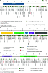
SNP mapping of methylenetetrahydrofolate reductase (MTHFR), methionine synthase (MTR), and methionine synthase reductase (MTRR). a The MTHFR protein depicted is a 656-amino acid protein (RefSeq: NP_005948, UniProt: P42898). The NAD(P)H domain that spans residues 48–337 was obtained from protein databases Pfam and InterPro. Alpha-helices are shown as green boxes; beta-strands as red triangles; FAD-binding sites as yellow circles; substrate binding sites as pink circles. b The MTR protein depicted is a 1265-amino acid protein (RefSeq: NP_000245.2, UniProt: Q99707). The Hcy-binding domain (residues 19–338), Pterin-binding domain (residues 371–632), B-12 binding domain (residues 662–907), and AdoMet activation domain (residues 923–1265) were obtained from protein database PROSITE. Alpha-helices are shown as green boxes; beta-strands as red triangles; zinc metal binding sites as blue circles; cobalt binding sites as yellow circles; cobalamin binding sites as pink circles; and S-adenosyl-l-methionine binding sites as purple circles. c The MTRR protein depicted is a 725 amino acid protein (RefSeq: NP_076915.2, UniProt: Q9UBK8). The flavodoxin-like domain (residues 32–174) and FAD-binding FR-type domain (residues 298–560) were obtained from protein database PROSITE (PS51384, PS50902). Alpha-helices are shown as green boxes; beta-strands as red triangles; NADP binding sites as yellow circles; and FAD-binding sites as pink circles. All predicted secondary structures were obtained from PSIPRED by the University College London Bioinformatics group (http://bioinf.cs.ucl.ac.uk/psipred). The binding sites were obtained from UniProt. Missense SNP IDs and locations were obtained from NCBI dbSNP. The DS-associated are shown in red
Fig. 3
Fig. 3
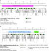
SNP mapping of reduced folate carrier 1 (RFC1) and cystathionine beta synthase (CBS). a The RFC1 protein depicted is a 591-amino acid protein (RefSeq: NP_919231, UniProt: P41440). The reduced folate carrier domain that spans residues 24–433 was obtained from protein databases Pfam (PF01770). Alpha-helices are shown as green boxes; beta-strands as red triangles. b The CBS protein depicted is a 551-amino acid protein (RefSeq: NP_000062.1, UniProt: P35520). The pyridoxal-phosphate dependent domain (residues 82–376) and CBS domain (residues 418–476) were obtained from protein database InterPro. Alpha-helices are shown as green boxes; beta-strands as red triangles; iron metal binding sites as yellow circles; pyridoxal phosphate binding sites as pink circles. The predicted secondary structure was obtained from PSIPRED by the University College London Bioinformatics group (http://bioinf.cs.ucl.ac.uk/psipred). Missense SNP IDs and locations were obtained from NCBI dbSNP. The DS-associated SNPs are shown in red
Fig. 4
Fig. 4
SNP mapping of p53. The p53 protein depicted is a 393-amino acid protein (RefSeq: NP_000537, UniProt: P04637). The transactivation domain (TAD) (residues 5–29), DNA-binding domain (residues 95–289) and tetramerization domain (residues 318–359) were obtained from the protein database Pfam. The predicted secondary structure was obtained from PSIPRED by the University College London Bioinformatics group (http://bioinf.cs.ucl.ac.uk/psipred). Alpha-helices are shown in green boxes; beta-strands as red triangles; DNA-interaction sites as yellow circles; zinc metal binding sites as purple circles. The binding sites were obtained from UniProt. Missense SNP IDs and locations were obtained from NCBI dbSNP. The DS-associated SNP (rs1042522) is shown in red

References

    1. Antonarakis SE, Petersen MB, McInnis MG, Adelsberger PA, Schinzel AA, Binkert F, et al. The meiotic stage of nondisjunction in Trisomy 21: determination by using DNA polymorphisms. Am J Hum Genet. 1992;50:544–550. - PMC - PubMed
    1. Canfield MA, Honein MA, Yuskiv N, Xing J, Mai CT, Collins JS, et al. National estimates and race/ethnic-specific variation of selected birth defects in the US, 1999–2001. Birth Defects Res A Clin Mol Teratol. 2006;76:747–756. doi: 10.1002/bdra.20294. - DOI - PubMed
    1. Bianca S. Non congenital heart disease aspects of Down’s syndrome. Images Paediatr Cardiol. 2002;4:3–11. - PMC - PubMed
    1. Fonatsch C. The role of chromosome 21 in hematology and oncology. Genes Chromosomes Cancer. 2010;49:497–508. - PubMed
    1. Antonarakis SE. Parental origin of the extra chromosome in Trisomy 21 as indicated by analysis of DNA polymorphisms. Down Syndrome Collaborative Group. N Engl J Med. 1991;324:872–876. doi: 10.1056/NEJM199103283241302. - DOI - PubMed

Publication types

MeSH terms