. 2016 Jun 1;13(1):132.
doi: 10.1186/s12974-016-0591-x.

Endogenous analgesia mediated by CD4(+) T lymphocytes is dependent on enkephalins in mice

Affiliations

Endogenous analgesia mediated by CD4(+) T lymphocytes is dependent on enkephalins in mice

Lilian Basso et al. J Neuroinflammation. .

Abstract

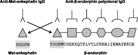
Background: T cell-derived opioids play a key role in the control of inflammatory pain. However, the nature of opioids produced by T cells is still matter of debate in mice. Whereas β-endorphin has been found in T lymphocytes by using antibody-based methods, messenger RNA (mRNA) quantification shows mainly mRNA encoding for enkephalins. The objective of the study is to elucidate the nature of T cell-derived opioids responsible for analgesia and clarify discrepancy of the results at the protein and genetic levels.

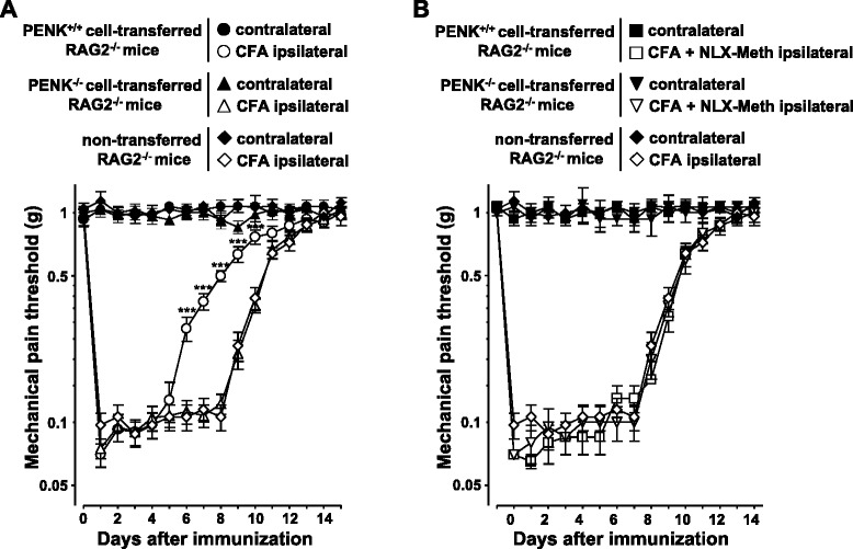
Methods: CD4(+) T lymphocytes were isolated from wild-type and enkephalin-deficient mice. mRNA encoding for β-endorphin and enkephalin was quantified by RT-qPCR. The binding of commercially available polyclonal anti-endorphin antibodies to lymphocytes from wild-type or enkephalin knockout mice was assessed by cytofluorometry. Opioid-mediated analgesic properties of T lymphocytes from wild-type and enkephalin-deficient mice were compared in a model of inflammation-induced somatic pain by measuring sensitivity to mechanical stimuli using calibrated von Frey filaments.

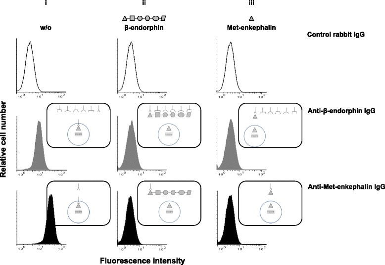
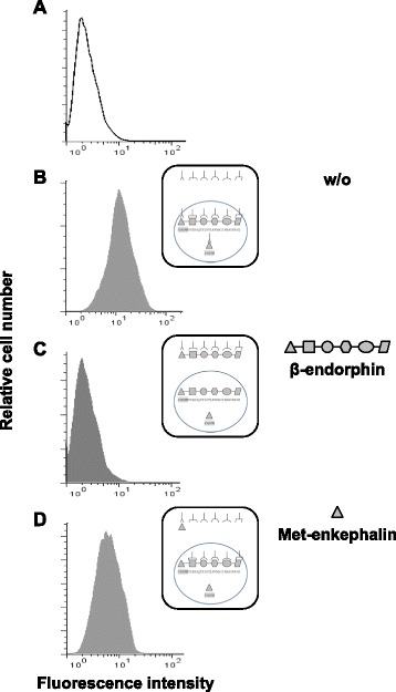
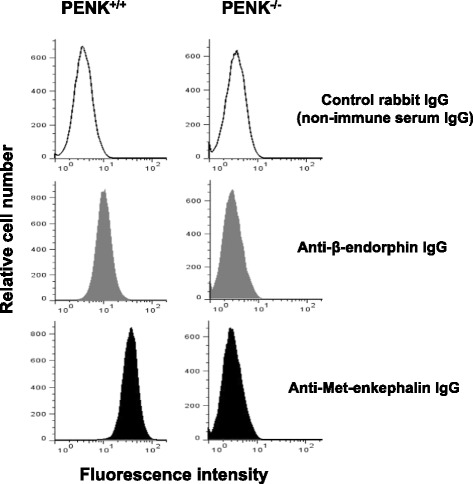
Results: CD4(+) T lymphocytes expressed high level of mRNA encoding for enkephalins but not for β-endorphin in mice. Anti-β-endorphin polyclonal IgG antibodies are specific for β-endorphin but cross-react with enkephalins. Anti-β-endorphin polyclonal antibodies bound to wild-type but not enkephalin-deficient CD4(+) T lymphocytes. Endogenous regulation of inflammatory pain by wild-type T lymphocytes was completely abolished when T lymphocytes were deficient in enkephalins. Pain behavior of immune-deficient (i.e., without B and T lymphocytes) mice was superimposable to that of mice transferred with enkephalin-deficient lymphocytes.

Conclusions: Rabbit polyclonal anti-β-endorphin serum IgG bind to CD4(+) T lymphocytes because of their cross-reactivity towards enkephalins. Thus, staining of T lymphocytes by anti-β-endorphin polyclonal IgG reported in most of studies in mice is because of their binding to enkephalins. In mice, CD4(+) T lymphocytes completely lose their analgesic opioid-mediated activity when lacking enkephalins.

Keywords: Enkephalin; Inflammation; Pain; T lymphocytes; β-endorphin.


Figures

Fig. 1
Fig. 1
Schematic representation of the potential cross-reactivity of anti-Met-enkephalin and anti-β-endorphin polyclonal IgG antibodies. The amino acid sequence of Met-enkephalin (five amino acids corresponding to only one epitope, gray box) is identical to the first five amino acids of the β-endorphin and may be recognized by immune serum IgG specifically raised against Met-enkephalin (formula image) or β-endorphin (formula image)
Fig. 2
Fig. 2
Proenkephalin is upregulated upon T cell activation. Naive CD4+ T lymphocytes isolated from wild-type mice were stimulated with both anti-CD3 and anti-CD28 mAbs for 6 days. Expression levels of mRNA encoding for PENK (gray histogram) and POMC (white histogram) were quantified by real-time PCR in CD4+ T lymphocytes from day 1 to day 6 (lower panels). mRNA content was normalized to the HPRT mRNA and quantified relative to standard mouse brain cDNA. Gene expression was assessed in at least five independent experiments run in duplicate. Results (mean ± SEM) are expressed relative to PENK or POMC mRNA expression in the mouse brain. Intracytoplasmic accumulation of Met-enkephalin-containing peptides was then assessed by cytofluorometry (upper panels). CD4+ T lymphocytes recovered each day of the stimulation were incubated with either control rabbit IgG (white histogram) or rabbit anti-Met-enkephalin IgG antibodies (gray histogram). The figure shows one representative experiment out of three performed
Fig. 3
Fig. 3
Anti-β-endorphin polyclonal IgG antibodies recognize enkephalins expressed in activated T lymphocytes as assessed by cytofluorometry. CD4+ T lymphocytes isolated from wild-type C57Bl/6 PENK+/+ mice were stimulated with both anti-CD3 and anti-CD28 mAbs for 6 days and intracellularly stained with control rabbit non-immune serum IgG (upper panels), rabbit anti-β-endorphin polyclonal IgG antibodies (middle panels), or rabbit anti-Met-enkephalin polyclonal IgG antibodies (lower panels). The figure depicts the binding of each of the three rabbit polyclonal IgG in the absence (i) or in the presence of an excess of soluble β-endorphin (ii) or Met-enkephalin (iii). The figure shows one representative experiment out of four performed. Schematic interpretation of the experiments is shown on the right of each histogram
Fig. 4
Fig. 4
The binding of anti-β-endorphin antibody to β-endorphin-expressing SK-N-MC cells is partially inhibited by soluble Met-enkephalin as assessed by cytofluorometry. The human neuroblastoma cell line SK-N-MC expressing both enkephalin and β-endorphin was intracellularly stained with control rabbit non-immune serum IgG (a) or rabbit anti-β-endorphin polyclonal IgG antibodies in the absence (b) or in the presence of an excess of soluble β-endorphin (c) or Met-enkephalin (d). The figure shows one representative experiment out of three performed. Schematic interpretation of the experiments is shown on the right of each histogram
Fig. 5
Fig. 5
Anti-β-endorphin polyclonal IgG antibodies do not bind to activated lymphocytes from pre-proenkephalin knockout mice as assessed by cytofluorometry. CD4+ T lymphocytes isolated from wild-type C57Bl/6 (PENK+/+) mice (left panels) or pre-proenkephalin knockout (PENK−/−) mice (right panels) were stimulated with both anti-CD3 and anti-CD28 mAbs for 6 days and intracellularly stained with either control rabbit non-immune serum IgG (upper panels), rabbit anti-β-endorphin polyclonal IgG antibodies (middle panels), or rabbit anti-Met-enkephalin polyclonal IgG antibodies (lower panels). The figure shows one representative experiment out of three performed
Fig. 6
Fig. 6
Anti-nociceptive T cell activity is mediated by enkephalins in mice. a Splenocytes from PENK+/+ wild-type C57Bl/6 (circle) or PENK−/− pre-proenkephalin knockout (triangle) mice were transferred i.v. into immune-deficient RAG-2−/− mice (n = 12 for each group of mice). The next day, mice were immunized s.c. into hind footpads with OVA in CFA. Immunized non-transferred RAG2−/− mice (n = 10, diamond) were used as control. Inflammatory pain was monitored by measuring sensitivity of the mice to mechanical stimuli using the von Frey test. Data are expressed as mean ± SEM paw withdrawal thresholds measured in inflamed ipsilateral immunized paws (open symbol) and contralateral non-injected paws (closed symbol). Results were obtained from two sets of experiments performed on groups of six (T cell transferred) or five (RAG2−/−) mice. ***p < 0.001 (comparison with immunized non-transferred RAG-2−/− mice). b From day 3 until the end of the experiment, recipient RAG2−/− mice transferred with either PENK+/+ (square; n = 6) or PENK−/− (inverted triangle; n = 6) splenocytes were daily injected into inflamed hind paw with 10 μL of naloxone methiodide (NLX-Meth) at 2 mg mL−1, 30 min before pain assessment. Mice were randomly assigned into each group in blind experiments in which the treatment and pain assessment were performed by two different experimenters. Data are expressed as mean ± SEM paw withdrawal thresholds measured in inflamed ipsilateral immunized paws (open symbol) and contralateral non-injected paws (closed symbol). Results were obtained from two sets of experiments performed on groups of mice mice

Similar articles

Cited by

References

    1. Boue J, Blanpied C, Brousset P, Vergnolle N, Dietrich G. Endogenous opioid-mediated analgesia is dependent on adaptive T cell response in mice. J Immunol. 2011;186:5078–84. doi: 10.4049/jimmunol.1003335. - DOI - PubMed
    1. Cabot PJ, Carter L, Gaiddon C, Zhang Q, Schäfer M, Loeffler J, Stein C. Immune cell-derived ß-endorphin. Production, release, and control of inflammatory pain in rats. J Clin Invest. 1997;100:142–8. doi: 10.1172/JCI119506. - DOI - PMC - PubMed
    1. Machelska H, Cabot PJ, Mousa SA, Zhang Q, Stein C. Pain control in inflammation governed by selectins. Nat Med. 1998;4:1425–8. doi: 10.1038/4017. - DOI - PubMed
    1. Machelska H, Schopohl JK, Mousa SA, Labuz D, Schafer M, Stein C. Different mechanisms of intrinsic pain inhibition in early and late inflammation. J Neuroimmunol. 2003;141:30–9. doi: 10.1016/S0165-5728(03)00213-3. - DOI - PubMed
    1. Rittner HL, Brack A, Machelska H, Mousa SA, Bauer M, Schafer M, Stein C. Opioid peptide-expressing leukocytes: identification, recruitment, and simultaneously increasing inhibition of inflammatory pain. Anesthesiology. 2001;95:500–8. doi: 10.1097/00000542-200108000-00036. - DOI - PubMed

Publication types