. 2016 Jun 1:6:26399.
doi: 10.1038/srep26399.

Rapid and sensitive detection of early esophageal squamous cell carcinoma with fluorescence probe targeting dipeptidylpeptidase IV

Affiliations

Rapid and sensitive detection of early esophageal squamous cell carcinoma with fluorescence probe targeting dipeptidylpeptidase IV

Haruna Onoyama et al. Sci Rep. .

Abstract

Early detection of esophageal squamous cell carcinoma (ESCC) is an important prognosticator, but is difficult to achieve by conventional endoscopy. Conventional lugol chromoendoscopy and equipment-based image-enhanced endoscopy, such as narrow-band imaging (NBI), have various practical limitations. Since fluorescence-based visualization is considered a promising approach, we aimed to develop an activatable fluorescence probe to visualize ESCCs. First, based on the fact that various aminopeptidase activities are elevated in cancer, we screened freshly resected specimens from patients with a series of aminopeptidase-activatable fluorescence probes. The results indicated that dipeptidylpeptidase IV (DPP-IV) is specifically activated in ESCCs, and would be a suitable molecular target for detection of esophageal cancer. Therefore, we designed, synthesized and characterized a series of DPP-IV-activatable fluorescence probes. When the selected probe was topically sprayed onto endoscopic submucosal dissection (ESD) or surgical specimens, tumors were visualized within 5 min, and when the probe was sprayed on biopsy samples, the sensitivity, specificity and accuracy reached 96.9%, 85.7% and 90.5%. We believe that DPP-IV-targeted activatable fluorescence probes are practically translatable as convenient tools for clinical application to enable rapid and accurate diagnosis of early esophageal cancer during endoscopic or surgical procedures.


Conflict of interest statement

The authors declare no competing financial interests.

Figures

Figure 1
Figure 1. Screening of HMRG-based aminopeptidase-reactive fluorescent probes.
(a) Activation of fluorescence of HMRG-based probes upon enzymatic reaction. (b) Fluorescence imaging of cancer-positive biopsy samples and cancer-negative biopsy samples. White-light image before spraying probe (left). Fluorescence images after spraying fluorescent probe under blue light (middle). Fluorescence images at 540 nm after spraying fluorescent probe; ROI are outlined in red (right). Tyr-HMRG was used as the fluorescent probe in these images. Scale bars, 5 mm. (c) Screening of aminopeptidase activity using human cancer-positive (SCC, solid line) and cancer-negative (N, dotted line) biopsy samples. Biopsy samples from the same patient are shown in the same color. F.I. (a.u.): fluorescence intensity (arbitrary units).
Figure 2
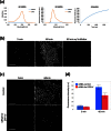
Figure 2. Fluorescence imaging of human esophageal squamous cell carcinoma cell line KYSE270 with GP-HMRG.
(a) In vitro changes in absorption (left) and fluorescence (middle) spectra of GP-HMRG before and after addition of DPP-IV. (b) Confocal fluorescence images at 5 and 60 min after incubation with GP-HMRG (10 μM) in the absence and presence of inhibitor (100 μM). Drastic fluorescence activation of GP-HMRG was observed, and was significantly suppressed by the inhibitor. Scale bars, 100 μm. (c) Confocal fluorescence images of KYSE270 cells which had been pretransfected with siRNAs, followed by application of GP-HMRG (10 μM). Scale bars, 100 μm. (d) Change of fluorescence intensity in KYSE270 cells in (b). Data are mean fluorescence intensities (a.u.) ± SEM.
Figure 3
Figure 3. Fluorescence analysis of freshly resected biopsy samples of ESCCs from patients.
Time-dependent changes in fluorescence intensity in biopsy samples after application of EP-HMRG (50 μM) (32 cancer-positive biopsy samples, red line; 42 cancer-negative biopsy samples, blue line). Mean fluorescence intensities (horizontal line within box), interquartile range (box) and range (error bars) are shown.
Figure 4
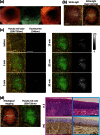
Figure 4. Fluorescence and histological analysis of freshly resected human ESCC specimen obtained at operation.
(a) White-light image (WLI) and lugol dye staining: the pathological diagnosis was 0-I + IIc, 85 mm in size, pT1b. Scale bar, 20 mm. (b) Fluorescence images after spraying EP-HMRG (50 μM) under blue light: a rapid fluorescence increase was observed at the tumor lesion. Scale bar, 20 mm. (c) Histological analysis of boxed regions with strong fluorescence activation (pink box) or with no fluorescence activation (blue box). H&E (upper) and IHC staining for DPP-IV (bottom) revealed that the pink box region consisted of SCCs with strong DPP-IV expression (middle) and the blue box region was normal (right). Magnification ×400. Scale bar, 100 μm.
Figure 5
Figure 5. Fluorescence and histological analysis of freshly resected ESD specimen.
(a) Endoscopic appearance. White light imaging (WLI) shows flat appearance and slight redness (left). Narrow band imaging (NBI) shows a brownish area (middle). Lugol dye shows a well-demarcated unstained area (right). (b) WLI and lugol staining: the pathological diagnosis was 0-IIb, 33 mm in size, pT1a. Scale bar, 10 mm. (c) Fluorescence images after spraying EP-HMRG (50 μM) under blue light: a rapid fluorescent increase was observed in the tumor lesion. Scale bar, 10 mm. (d) Red lines on histological mapping show SCCs and blue lines show no tumor (left). Histological analysis of boxed regions with strong fluorescence activation (pink box) or with no fluorescence activation (blue box). H&E (upper) and IHC staining for DPP-IV (bottom) revealed the pink box region was SCCs (middle) and the blue box region was normal (right). Magnification ×400. Scale bar, 100 μm.

References

    1. Enzinger P. C. & Mayer R. J. Esophageal cancer. N Engl J Med. 349, 2241–2252 (2003). - PubMed
    1. Katada C. et al.. Clinical outcome after endoscopic mucosal resection for esophageal squamous cell carcinoma invading the muscularis mucosae–a multicenter retrospective cohort study. Endoscopy. 39, 779–783 (2007). - PubMed
    1. Katada C., Muto M., Manabe T., Ohtsu A. & Yoshida S. Local recurrence of squamous-cell carcinoma of the esophagus after EMR. Gastrointest Endosc. 61, 219–225 (2005). - PubMed
    1. Shimizu Y. et al.. Endoscopic submucosal dissection for treatment of early stage hypopharyngeal carcinoma. Gastrointest Endosc. 64, 255–259 (2006). - PubMed
    1. Ono S. et al.. Long-term outcomes of endoscopic submucosal dissection for superficial esophageal squamous cell neoplasms. Gastrointest Endosc. 70, 860–866 (2009). - PubMed

Publication types

MeSH terms

Associated data