

doi: 10.1007/978-1-4939-3603-8_26.
AOM/DSS Model of Colitis-Associated Cancer
Affiliations
- PMID: 27246042
- PMCID: PMC5035391
- DOI: 10.1007/978-1-4939-3603-8_26
Item in Clipboard
AOM/DSS Model of Colitis-Associated Cancer
Methods Mol Biol.
2016.
Abstract
Our understanding of colitis-associated carcinoma (CAC) has benefited substantially from mouse models that faithfully recapitulate human CAC. Chemical models, in particular, have enabled fast and efficient analysis of genetic and environmental modulators of CAC without the added requirement of time-intensive genetic crossings. Here we describe the Azoxymethane (AOM)/Dextran Sodium Sulfate (DSS) mouse model of inflammatory colorectal cancer.
Keywords: AOM; Colitis-associated cancer; Colon cancer; DSS; Inflammatory carcinogenesis.
Figures
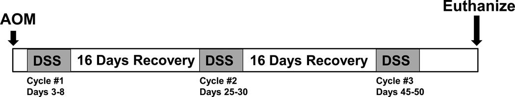
Schematic timeline for AOM/DSS induced inflammatory carcinogenesis.

Example of weight loss during repeat cycle DSS treatment.
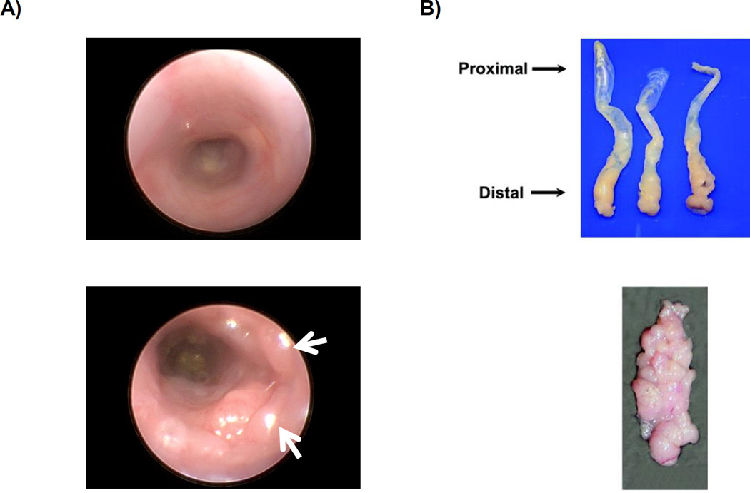
A) Above: Endoscopy image of normal colon. Below: Endoscopy of colon after AOM injection followed by two cycles of DSS. White arrows indicate tumors. B) Above: Colons harvested and oriented with the distal end toward the dissector. Below: Example of gross tumor burden. Images are reproduced from prior report (23).
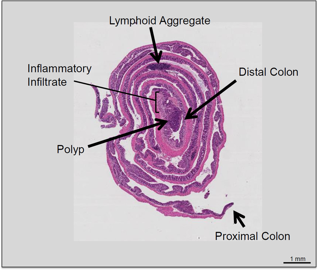
Hematoxylin and eosin stain of Swiss-rolled colon. The most distal portion of the colon is located in the innermost segment of the roll. A large polyp can be seen in the distal colon surrounded by inflammatory infiltrate.
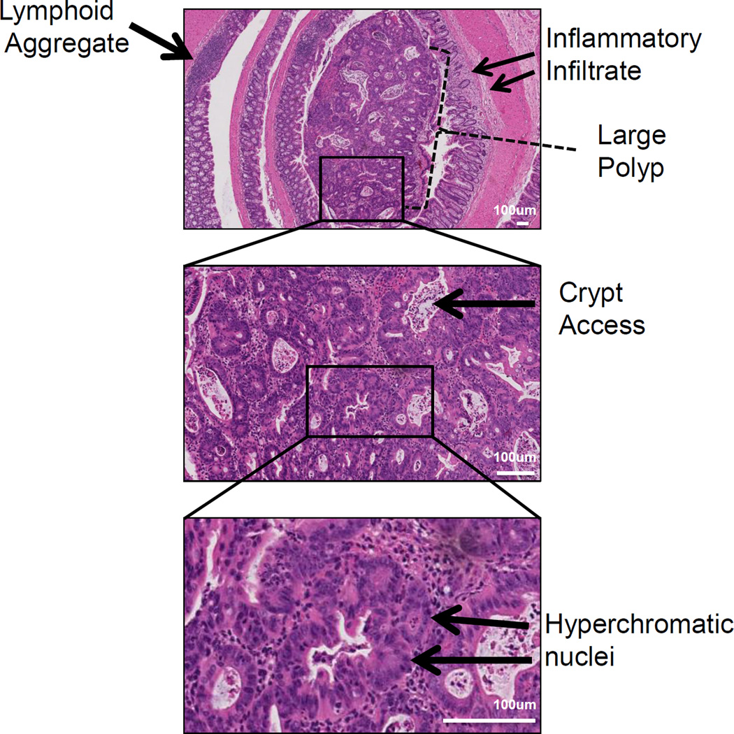
Representative histology of a distal colonic tumor isolated from a mouse treated with the AOM/DSS protocol. Pathology features indicating injury include inflammatory infiltrates are observed at low power (top panel) and intratumoral crypt abscesses (middle panel). Features of neoplasia such as hyperchromatic nuclei and increased nuclei/cytoplasmic ratios are identified at higher magnification (middle and bottom panel).
References
-
- Siegel R, Ma J, Jemal A. Cancer statistics, 2014. CA. Cancer J Clin. 2014;64:9–29. - PubMed
-
- Danese S, Malesci A, Vetrano S. Colitis-associated cancer: the dark side of inflammatory bowel disease. Gut. 2011;60:1609–1610. - PubMed
-
- Danese S an, Mantovani A. Inflammatory bowel disease and intestinal cancer : a paradigm of the Yin – Yang interplay between inflammation and cancer. Oncogene. 2010;29:3313–3323. - PubMed
-
- Terzić J, Grivennikov S, Karin E, Karin M. Inflammation and colon cancer. Gastroenterology. 2010;138:2101–2114. - PubMed
Publication types
MeSH terms
Substances
Grants and funding
LinkOut - more resources
Full Text Sources
Other Literature Sources
Medical
