. 2016 Jul;139(Pt 7):2002-14.
doi: 10.1093/brain/aww118. Epub 2016 May 30.

Transcranial magnetic stimulation and potential cortical and trigeminothalamic mechanisms in migraine

Affiliations

Transcranial magnetic stimulation and potential cortical and trigeminothalamic mechanisms in migraine

Anna P Andreou et al. Brain. 2016 Jul.

Abstract

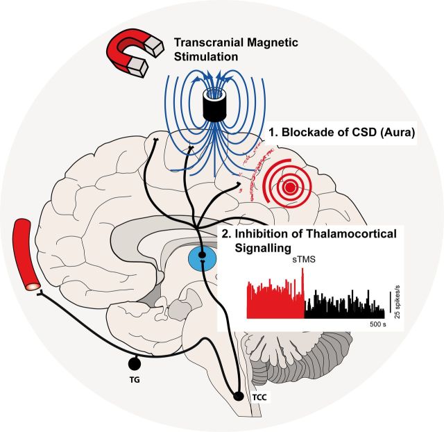
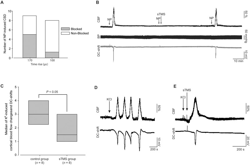
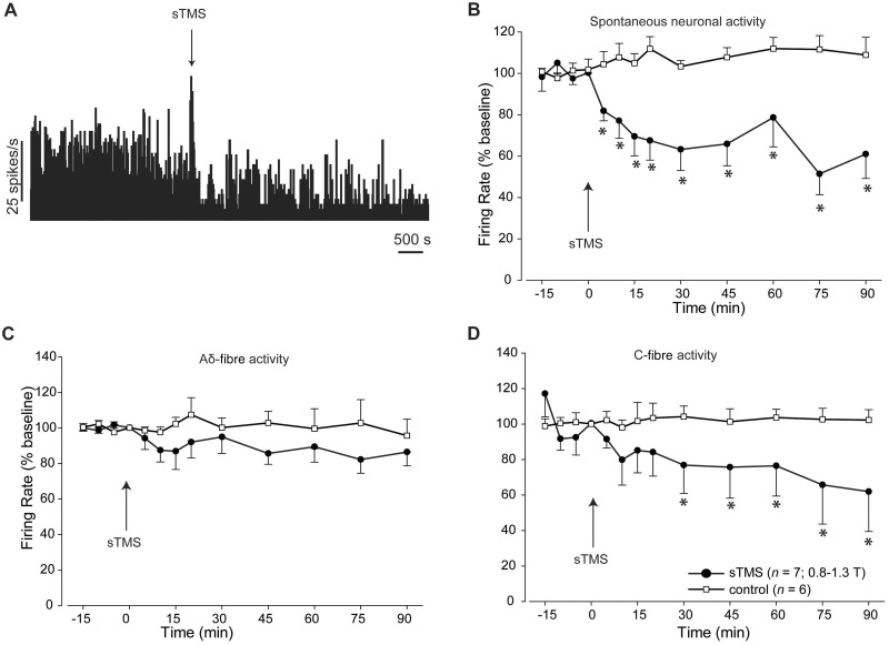
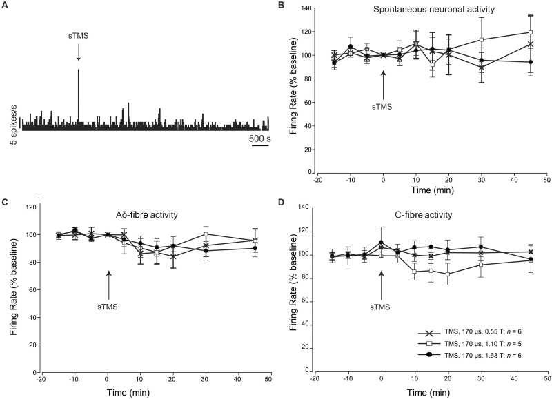
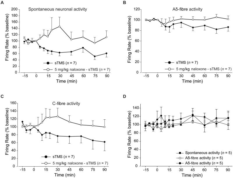
A single pulse of transcranial magnetic stimulation has been shown to be effective for the acute treatment of migraine with and without aura. Here we aimed to investigate the potential mechanisms of action of transcranial magnetic stimulation, using a transcortical approach, in preclinical migraine models. We tested the susceptibility of cortical spreading depression, the experimental correlate of migraine aura, and further evaluated the response of spontaneous and evoked trigeminovascular activity of second order trigemontothalamic and third order thalamocortical neurons in rats. Single pulse transcranial magnetic stimulation significantly inhibited both mechanical and chemically-induced cortical spreading depression when administered immediately post-induction in rats, but not when administered preinduction, and when controlled by a sham stimulation. Additionally transcranial magnetic stimulation significantly inhibited the spontaneous and evoked firing rate of third order thalamocortical projection neurons, but not second order neurons in the trigeminocervical complex, suggesting a potential modulatory effect that may underlie its utility in migraine. In gyrencephalic cat cortices, when administered post-cortical spreading depression, transcranial magnetic stimulation blocked the propagation of cortical spreading depression in two of eight animals. These results are the first to demonstrate that cortical spreading depression can be blocked in vivo using single pulse transcranial magnetic stimulation and further highlight a novel thalamocortical modulatory capacity that may explain the efficacy of magnetic stimulation in the treatment of migraine with and without aura.

Keywords: aura; cortical spreading depression; migraine; thalamus; transcranial magnetic stimulation.


Figures

None
Migraine affects 1 billion people worldwide, and is the most common cause of neurological disability. Single pulse transcranial magnetic stimulation (sTMS) has been shown to be effective for the acute treatment of migraine. Using preclinical models, Andreou, Holland et al . explore thalamocortical mechanisms as a basis for this clinical effect.
Figure 1
Figure 1
Experimental setup and design. ( A ) Experimental set-up in the rat CSD model. Cortical steady potential (DC-shift) via a Ag/AgCl glass microelectrode was recorded in addition to cerebral blood flow changes from a single laser Doppler probe, ∼2 mm anterior to lamda. CSD was induced at the occipital cortex either mechanically (needle prick) or chemically (KCl). A single pulse TMS (sTMS) coil was positioned 5–7 mm above the cortex, close to the CSD induction site. ( B ) Experimental set-up in cat CSD model. CSD was monitored via cerebral blood flow changes, measured via two laser Doppler probes; probe 1 was placed a few millimetres rostral to the CSD induction side in the occipital cortex. Probe 2 was placed in the ipsilateral frontal cortex, a few millimetres anterior to the bregma line. A single pulse TMS coil was positioned 5 mm above the cortex, close to the CSD induction site. ( C ) Experimental set-up in the trigeminovascular activation model for recordings from the TCC. Extracellular electrophysiological recordings were performed from second order neurons in the TCC that demonstrated stable activation in response to trigeminovascular stimulation. A bipolar stimulating electrode onto the middle meningeal artery (MMA) provided square-pulse electrical stimulation for trigeminovascular activation. A single pulse TMS coil was positioned 5 mm above the occipital cortex. ( D ) Experimental set-up in the trigeminovascular activation model for recordings from the ventroposteromedial thalamic nucleus (VPM). Extracellular electrophysiological recordings were performed from third order neurons in the VPM, following stereotaxic positioning of a recording electrode (through the parietal cortex). A bipolar stimulating electrode onto the superior sagittal sinus (SSS) provided square-pulse electrical stimulation for trigeminovascular activation. A single pulse TMS coil was positioned ∼5–7 mm above the occipital cortex. ( E ) Bespoke interchangeable Kopf mountable single pulse TMS coil, kept in an insulated cup of 30 mm diameter. ( F ) A representative magnetic pulse and pulse characteristics, as recorded in a data management system and displayed on an oscilloscope, through a pulse detection circuit. ( G ) A diagram illustrating the experimental protocol employed in the mechanically induced CSD models (cats and rats). ( H ) A diagram illustrating the experimental protocol used for the K + -induced CSD model. ( I ) A diagram illustrating the experimental protocol used for the trigeminovascular activation models (for both TCC and VPM recordings). In a group of animals used for trigeminovascular activation and recordings in the VPM, naloxone was used as a pretreatment to single pulse TMS.
Figure 2
Figure 2
The impact of TMS on mechanically and chemically induced CSD. ( A ) Single pulse TMS (sTMS) delivered though a coil with rise time of 170 µs (1.11–1.63 T) significantly blocked mechanically (needle prick, NP) induced CSDs in five of nine rats when pulsed 30 s post CSD induction. Single pulse TMS delivered though a coil with rise time of 100 µs, inhibited mechanically induced CSD on the rat cortex in one of eight rats. ( B ) Representative example of a CSD blockade by single pulse TMS stimulation. Example demonstrates the successful induction of CSD pre-single pulse TMS and recovery post-blockade by single pulse TMS. Animals blood pressure recordings were unaffected by single pulse TMS. ( C ) Single pulse TMS applied over the rat cortex using a coil of a rise time of 170 µs significantly decreased the frequency of CSD waves, measured by the number of CBF and intracortical DC-shift changes within 60 min after topical potassium chloride (KCl) application. ( D ) Representative example of CSD waves induced by topical KCl application in an untreated animal. ( E ) Representative example of CSD waves induced by topical KCl application in an animal treated by single pulse TMS (170 µs, 1.1 T) 30 min post-KCl application. CBF = cerebral blood flow.
Figure 3
Figure 3
The impact of TMS on thalamocoritcal neurons in the ventroposteromedial thalamic nucleus. ( A ) Representative example demonstrating the effect of single pulse TMS on spontaneous neuronal activity discriminated from third order neurons recorded in the ventroposteromedial thalamic nucleus. A characteristic artefact was recorded during single pulse TMS. ( B ) Single pulse TMS significantly reduced the spontaneous neuronal activity recorded from third order neurons in the ventroposteromedial thalamus. ( C and D ) Single pulse TMS had no significant effect on the evoked trigeminovascular activity recorded from third order neurons in the ventroposteromedial thalamus in response to Aδ-fibres ( C ); however, it significantly inhibited evoked trigeminovascular activity in response to C-fibre activation ( D ). In the control group ( n = 6), in which no single pulse TMS pulse was delivered, evoked and spontaneous neuronal activity from third order neurons was recorded in the absence of any single pulse TMS application.
Figure 4
Figure 4
The impact of TMS on trigeminothalamic projection neurons in the TCC. ( A ) Representative example demonstrating the effect of single pulse TMS (sTMS) on spontaneous neuronal activity discriminated from second order neurons recorded in the TCC. A characteristic artefact was recorded during single pulse TMS. ( B ) Single pulse TMS applied with a coil of 170 µs rise time at different tesla intensities, had no significant effect on the spontaneous neuronal activity recorded from second order neurons in the TCC. ( C and D ) Single pulse TMS applied with a coil of 170 µs rise time at different tesla intensities, had no significant effect on the evoked trigeminovascular activity recorded from second order neurons in the TCC, in response to Aδ- ( C ) and C-fibre ( D ) Neuronal activity was followed for 90 min post-single pulse TMS; however, as no significant effect occurred, graphs in B–D report data for the first 45 min post-single pulse TMS.
Figure 5
Figure 5
The impact of opiodergic mechanisms on thalamocortical TMS responses. Pretreatment with naloxone (5 mg/kg; intravenous bolus) 5 min pre-single pulse TMS application significantly blocked the inhibitory effects of single pulse TMS over the spontaneous and evoked firing of third order neurons. Graphs demonstrate the comparison between the single pulse TMS group with and without naloxone pre-treatment over the spontaneous neuronal activity ( A ) and trigeminovascular evoked activity in response to Aδ- ( B ) and C-fibre activation ( C ). ( D ) Naloxone administered alone, in the absence of single pulse TMS, induced no significant changes over spontaneous or evoked trigeminovascular activity in response to Aδ- and C-fibre activation.

References

    1. Afridi SK, Goadsby PJ . Neuroimaging of migraine . Curr Pain Headache Rep 2006. ; 10 : 221 – 4 . - PubMed
    1. Akerman S, Holland P, Goadsby PJ . Diencephalic and brainstem mechanisms in migraine . Nat Rev Neurosci 2011. ; 12 : 570 – 84 . - PubMed
    1. Akerman S, Holland PR, Goadsby PJ . Mechanically-induced cortical spreading depression associated regional cerebral blood flow changes are blocked by Na+ ion channel blockade . Brain Res 2008. ; 1229 : 27 – 36 . - PubMed
    1. Akerman S, Holland PR, Lasalandra M, Goadsby PJ . Endocannabinoids in the brainstem modulate dural trigeminovascular nociceptive traffic via CB1 and ‘triptan' receptors: implications in migraine . J Neurosci 2013. ; 33 : 14869 – 77 . - PMC - PubMed
    1. Ambriz-Tututi M, Sanchez-Gonzalez V, Drucker-Colin R . Transcranial magnetic stimulation reduces nociceptive threshold in rats . J Neurosci Res 2012. ; 90 : 1085 – 95 . - PubMed

Publication types