. 2016 Jul 28;198(16):2156-65.
doi: 10.1128/JB.00101-16. Print 2016 Aug 15.

Rotation of Vibrio fischeri Flagella Produces Outer Membrane Vesicles That Induce Host Development

Affiliations

Rotation of Vibrio fischeri Flagella Produces Outer Membrane Vesicles That Induce Host Development

Marie-Stephanie Aschtgen et al. J Bacteriol. .

Abstract

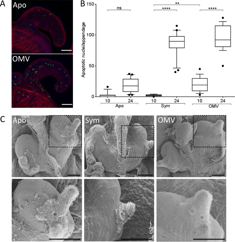
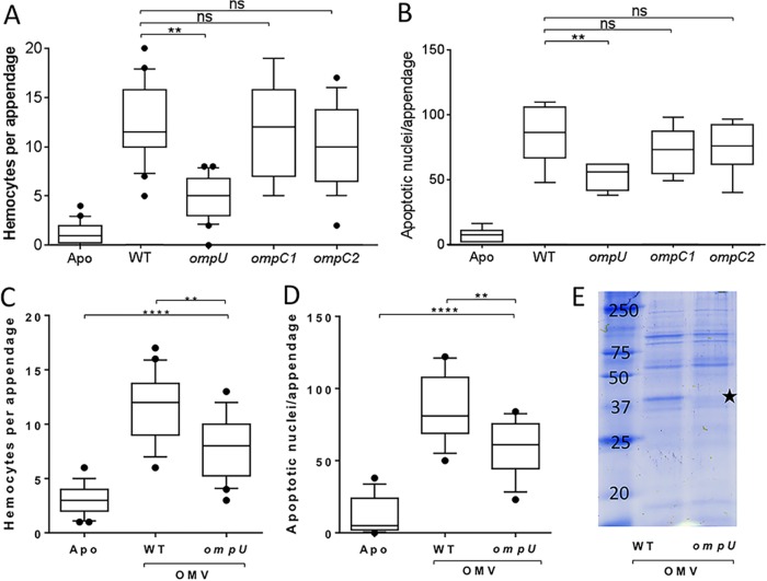
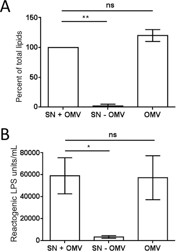
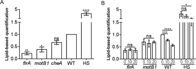
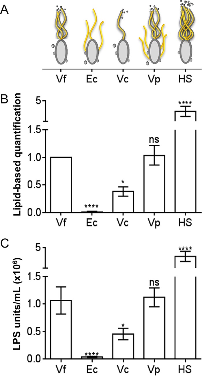
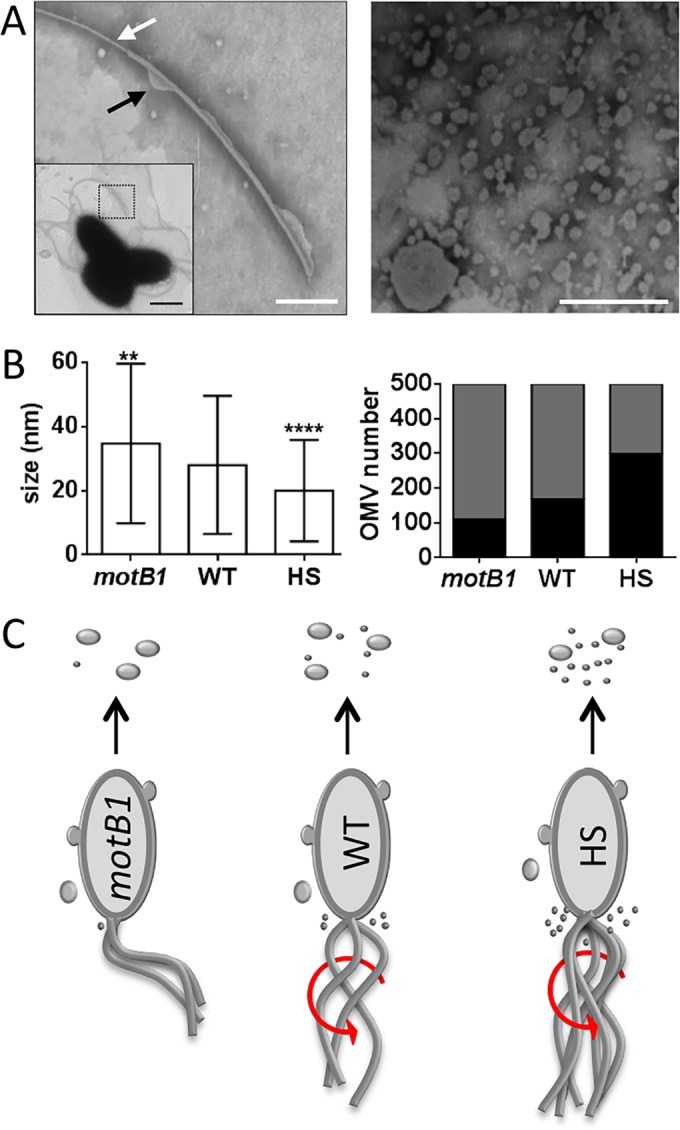
Using the squid-vibrio association, we aimed to characterize the mechanism through which Vibrio fischeri cells signal morphogenesis of the symbiotic light-emitting organ. The symbiont releases two cell envelope molecules, peptidoglycan (PG) and lipopolysaccharide (LPS) that, within 12 h of light organ colonization, act in synergy to trigger normal tissue development. Recent work has shown that outer membrane vesicles (OMVs) produced by V. fischeri are sufficient to induce PG-dependent morphogenesis; however, the mechanism(s) of OMV release by these bacteria has not been described. Like several genera of both beneficial and pathogenic bacteria, V. fischeri cells elaborate polar flagella that are enclosed by an extension of the outer membrane, whose function remains unclear. Here, we present evidence that along with the well-recognized phenomenon of blebbing from the cell's surface, rotation of this sheathed flagellum also results in the release of OMVs. In addition, we demonstrate that most of the development-inducing LPS is associated with these OMVs and that the presence of the outer membrane protein OmpU but not the LPS O antigen on these OMVs is important in triggering normal host development. These results also present insights into a possible new mechanism of LPS release by pathogens with sheathed flagella.

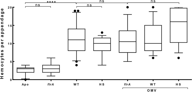
Importance: Determining the function(s) of sheathed flagella in bacteria has been challenging, because no known mutation results only in the loss of this outer membrane-derived casing. Nevertheless, the presence of a sheathed flagellum in such host-associated genera as Vibrio, Helicobacter, and Brucella has led to several proposed functions, including physical protection of the flagella and masking of their immunogenic flagellins. Using the squid-vibrio light organ symbiosis, we demonstrate another role, that of V. fischeri cells require rotating flagella to induce apoptotic cell death within surface epithelium, which is a normal step in the organ's development. Further, we present evidence that this rotation releases apoptosis-triggering lipopolysaccharide in the form of outer membrane vesicles. Such release may also occur by pathogens but with different outcomes for the host.


Figures

FIG 1
FIG 1
OMVs induce phenotypes associated with LPS-linked light organ morphogenesis. (A) Representative Z-stack confocal images of TUNEL-stained aposymbiotic juvenile light organs, with (OMV) or without (Apo) the addition of OMVs. Filamentous actin was stained with rhodamine-633 (red), nuclei were stained with TOTO-3 (blue), and apoptotic nuclei were TUNEL stained (green). Scale bars = 50 μm. (B) Induction of apoptosis in the superficial epithelium. Animals were untreated (Apo), exposed to 10,000 CFU V. fischeri/ml (Sym), or exposed to 50 μg of OMV protein/ml (OMV) for either 10 or 24 h. Apoptotic nuclei were counted in one appendage per light organ. Error bars indicate one-way ANOVA (F = 184); here and elsewhere, the horizontal lines in the boxes indicate the median values and the boxes indicate the limits of the first and third quartiles. ****, P < 0.0001; **, P < 0.01; ns, not significant. (C) Two levels of scanning electron microscopy magnification (each image on the bottom is a magnification of the image in the dashed box above it) of the ventral light organ surfaces of representative 4-day-old animals, revealing the degree of ciliated epithelial field regression. Scale bars = 100 μm. Aposymbiotic animals (Apo) show an intact ciliated ridge, and both the anterior and posterior appendages are present and functional. Animals treated with 50 μg OMV protein/ml (OMV) show a level of regression similar to that of symbiotic animals (Sym).
FIG 2
FIG 2
OmpU is required for induction of two symbiotic phenotypes. (A and C) Levels of hemocyte trafficking in the anterior appendages of animals after 18 h. (B and D) Counts of apoptotic nuclei in the anterior appendages of animals after 24 h. (A and B) Animals were exposed to no V. fischeri cells (Apo), wild-type V. fischeri (WT), or one of three OMP mutants: the ompU mutant, the ompC1 mutant, or the ompC2 mutant. (C and D) Animals were exposed to OMVs (50 μg of OMV protein/ml) released by either the wild-type (WT) or the ompU mutant. One-way ANOVA was carried out (F = 31, 54, 39, and 15 for panels A, B, C, and D, respectively). ****, P < 0.0001; **, P < 0.01; ns, not significant. (E) Coomassie-stained SDS-PAGE gel of the proteins of OMVs released by either the wild-type (WT) or the ompU mutant (5 μg of OMV protein was loaded in each well). The star indicates the migration position of OmpU.
FIG 3
FIG 3
Lipid and LPS are released almost entirely as OMVs. OMVs were prepared from cell-free cultures of V. fischeri. Both total lipids and LPS were quantified in three fractions: culture supernatant (SN + OMV), the supernatant after OMV removal (SN − OMV), and the pelleted OMVs (OMV). (A) FM4-64-based total-lipid assay. (B) Limulus amoebocyte lysate (LAL) assay for LPS. A one-way repeated-measures ANOVA with a post hoc Bonferroni correction was performed. **, P < 0.01; *, P < 0.1; ns, not significant.
FIG 4
FIG 4
Flagellar rotation and motility promote OMV production. (A) Fold difference, relative to the wild type (WT), in the level of OMV production by four motility mutants: the flrA mutant (no flagellum), the motB1 mutant (nonrotating flagellum), the cheA mutant (chemotaxis defect), and a hyperflagellated swarmer mutant (HS). (B) Influence of medium viscosity (i.e., addition of 0, 10, or 20% Percoll) on relative levels of OMV production. Values are means ± standard errors of the means (SEM; n = 3). ****, P < 0.0001; ***, P < 0.001; **, P < 0.01; *, P < 0.1; ns, not significant.
FIG 5
FIG 5
Presence of sheathed flagella correlates with OMV release. (A) Cartoon comparing different bacterial strains, their flagella (yellow), and the presence of a flagellar sheath (gray). Vf, V. fischeri; Ec, E. coli; Vc, V. cholerae; Vp, V. parahaemolyticus; HS, a hyperflagellated swarmer V. fischeri mutant. (B and C) Fold difference, relative to V. fischeri, in OMV production. (B) Total-lipid-based assay for OMVs. (C) LPS-based assay for OMVs. A one-way repeated-measures ANOVA with a post hoc Bonferroni correction was performed. ****, P < 0.0001; *, P < 0.1; ns, not significant.
FIG 6
FIG 6
Flagellar activity is associated with reduced OMV size. (A) Left, negative-stained transmission electron micrograph (TEM) enlargement of a V. fischeri sheathed flagellum; white arrow, area exposing flagellar filament; black arrow, bleb forming on sheath; scale bar = 250 nm. Inset, TEM of three cells, with the flagellum in the area of enlargement (dotted box); scale bar = 1 μm. Right, TEM image of an OMV preparation; scale bar = 200 nm. (B) Left, mean size of OMVs produced by wild-type cells (WT) or by immotile mutant (motB1) or hyperflagellated swarmer (HS) derivatives. Right, proportion of OMVs whose size is less than (black bar) or greater than (gray bar) 20 nm. Error bars in the left panel indicate SEM (n = 500). ****, P < 0.0001; **, P < 0.01 (t test). (C) Cartoon depicting OMV production by different V. fischeri strains; the presence of sheathed flagella and their ability to rotate (red arrows), as well as the relative proportion of OMV sizes released, are illustrated.
FIG 7
FIG 7
Induction of hemocyte trafficking by OMVs released by motility mutants. Levels of hemocyte trafficking in the anterior appendages of animals were determined 18 h after exposure to cells or OMVs. Treatment was with no V. fischeri cells (Apo), wild-type V. fischeri (WT), or motility mutants lacking either flagella (flrA) or hyperflagella (HS). Animals were also exposed to OMVs (50 μg of OMV protein/ml) released by the three strains. One-way ANOVA, F = 22. ****, P < 0.0001; ns, not significant.

Similar articles

Cited by

References

    1. Ruby EG. 2008. Symbiotic conversations are revealed under genetic interrogation. Nat Rev Microbiol 6:752–762. doi:10.1038/nrmicro1958. - DOI - PMC - PubMed
    1. McFall-Ngai M, Heath-Heckman EAC, Gillette AA, Peyer SM, Harvie EA. 2012. The secret languages of coevolved symbioses: insights from the Euprymna scolopes-Vibrio fischeri symbiosis. Semin Immunol 24:3–8. doi:10.1016/j.smim.2011.11.006. - DOI - PMC - PubMed
    1. Koropatnick TA, Kimbell JR, McFall-Ngai MJ. 2007. Responses of host hemocytes during the initiation of the squid-Vibrio symbiosis. Biol Bull 212:29–39. doi:10.2307/25066578. - DOI - PubMed
    1. Kremer N, Philipp EER, Carpentier M-C, Brennan CA, Kraemer L, Altura MA, Augustin R, Häsler R, Heath-Heckman EAC, Peyer SM, Schwartzman J, Rader BA, Ruby EG, Rosenstiel P, McFall-Ngai MJ. 2013. Initial symbiont contact orchestrates host-organ-wide transcriptional changes that prime tissue colonization. Cell Host Microbe 14:183–194. doi:10.1016/j.chom.2013.07.006. - DOI - PMC - PubMed
    1. Montgomery MK, McFall-Ngai M. 1994. Bacterial symbionts induce host organ morphogenesis during early postembryonic development of the squid Euprymna scolopes. Development 120:1719–1729. - PubMed

Substances

LinkOut - more resources