. 2016 May 31;14(1):153.
doi: 10.1186/s12967-016-0907-8.

Cathepsin A inhibition attenuates myocardial infarction-induced heart failure on the functional and proteomic levels

Affiliations

Cathepsin A inhibition attenuates myocardial infarction-induced heart failure on the functional and proteomic levels

Agnese Petrera et al. J Transl Med. .

Abstract

Background: Myocardial infarction (MI) is a major cause of heart failure. The carboxypeptidase cathepsin A is a novel target in the treatment of cardiac failure. We aim to show that recently developed inhibitors of the protease cathepsin A attenuate post-MI heart failure.

Methods: Mice were subjected to permanent left anterior descending artery (LAD) ligation or sham operation. 24 h post-surgery, LAD-ligated animals were treated with daily doses of the cathepsin A inhibitor SAR1 or placebo. After 4 weeks, the three groups (sham, MI-placebo, MI-SAR1) were evaluated.

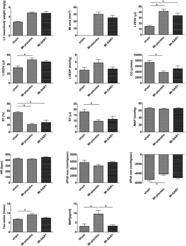
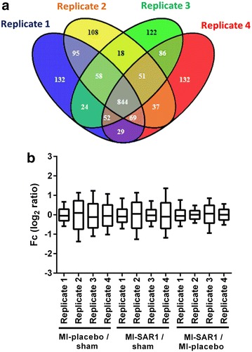
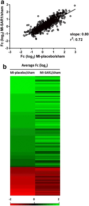
Results: Compared to sham-operated animals, placebo-treated mice showed significantly impaired cardiac function and increased plasma BNP levels. Cathepsin A inhibition prevented the increase of plasma BNP levels and displayed a trend towards improved cardiac functionality. Proteomic profiling was performed for the three groups (sham, MI-placebo, MI-SAR1). More than 100 proteins were significantly altered in placebo-treated LAD ligation compared to the sham operation, including known markers of cardiac failure as well as extracellular/matricellular proteins. This ensemble constitutes a proteome fingerprint of myocardial infarction induced by LAD ligation in mice. Cathepsin A inhibitor treatment normalized the marked increase of the muscle stress marker CA3 as well as of Igγ 2b and fatty acid synthase. For numerous further proteins, cathepsin A inhibition partially dampened the LAD ligation-induced proteome alterations.

Conclusions: Our proteomic and functional data suggest that cathepsin A inhibition has cardioprotective properties and support a beneficial effect of cathepsin A inhibition in the treatment of heart failure after myocardial infarction.

Keywords: Cardiovascular diseases; Drug therapy; Heart failure; Mouse model; Myocardial infarction.


Figures

Fig. 1
Fig. 1
Experimental design of the study. Sham operation or permanent LAD ligation were performed in mice. LAD ligation was either treated with placebo or with the cathepsin A inhibitor SAR1. After 28 days cardiac functionality was evaluated and a quantitative proteome study was conducted
Fig. 2
Fig. 2
Functional parameters in the murine MI model. Cardiac parameters were assessed 4 weeks after permanent ligation of the LAD. Three groups have been compared: sham (n = 10), MI-placebo (n = 15), MI-SAR1 (n = 10). Results are expressed as mean ± SEM. Differences significant at p < 0.05 are marked with an asterisk (*). LVESV left ventricular end-systolic volume, LVEDV left ventricular end-diastolic volume, LVEDP left ventricular end-diastolic pressure, CO cardiac output, EF ejection fraction, SV stroke volume, MAP mean arterial pressure, HR heart rate, dP/dt peak positive or negative first derivative of LV pressure, BNP plasma brain natriuretic peptide
Fig. 3
Fig. 3
Protein identification and quantification overview for the proteome analysis of murine hearts. a Venn diagram representing the number of identified proteins in each replicate and their overlap. 1074 proteins were found in at least three replicates. b Distribution and geometric mean (horizontal bar) of fold change values (log2 of relative protein ratios) of proteins from the four biological replicates. Each replicate contains an animal from each group (sham, MI-placebo, MI-SAR1)
Fig. 4
Fig. 4
Cardiac proteome fingerprint of cathepsin A inhibition upon LAD ligation. a Scatter plot and linear regression analysis of protein abundance alterations observed for placebo-treated LAD ligation (as compared to sham operation) and SAR1-treated LAD ligation (as compared to sham operation). b Heatmap comparison of fold changes MI-placebo/sham and MI-SAR1/sham of the 104 significantly affected proteins upon LAD ligation. Fold changes in the first column (MI-placebo/sham) are placed in descending order
Fig. 5
Fig. 5
Effect of cathepsin A inhibition on proteome alterations in LAD ligation. a Bar chart depiction of proteins that are significantly increased in placebo-treated LAD ligation (as compared to sham operation) and for which SAR1 treatment decreased this effect by a fold-change value of more than 50 % [difference of log2 ratios (MI-placebo/sham)–(MI-SAR1/sham) > 0.58]. b Same as (a) but for significantly decreased proteins in LAD ligation. Results are expressed as mean ± SEM. Differences significant at p < 0.05 are marked with an asterisk (*). c Western blot analysis of selected proteins. Shown here is a representative blot. GAPDH is used as loading control
Fig. 6
Fig. 6
Effect of cathepsin A inhibition in an in vitro model of ischemia. Cells were incubated for 24 h with 10 μM SAR1 or DMSO as a solvent-only control in simulated ischemia culture conditions (hypoxia and serum deprivation, see Methods section for details). a Quantitative analysis of caspase-3 activity in the total cell lysate of cells treated with SAR1 in standard conditions (normoxia; DMEM supplemented with FCS) and simulated ischemia (hypoxia; DMEM serum-free). b Cells in simulated ischemia were stained with YO-PRO-1 or PI and evaluated by flow cytometry. The percentage of cells positive to YO-PRO-1 or to PI was calculated. Results are expressed as mean ± SEM. Differences significant at p < 0.05 are marked with an asterisk (*)

References

    1. Bonten EJ, Annunziata I, d’Azzo A. Lysosomal multienzyme complex: pros and cons of working together. Cell Mol Life Sci. 2014;71:2017–2032. doi: 10.1007/s00018-013-1538-3. - DOI - PMC - PubMed
    1. Hiraiwa M. Cathepsin A/protective protein: an unusual lysosomal multifunctional protein. Cell Mol Life Sci. 1999;56:894–907. doi: 10.1007/s000180050482. - DOI - PMC - PubMed
    1. Seyrantepe V, Hinek A, Peng J, Fedjaev M, Ernest S, Kadota Y, et al. Enzymatic activity of lysosomal carboxypeptidase (cathepsin) A is required for proper elastic fiber formation and inactivation of endothelin-1. Circulation. 2008;117:1973–1981. doi: 10.1161/CIRCULATIONAHA.107.733212. - DOI - PubMed
    1. Petrera A, Lai ZW, Schilling O. Carboxyterminal protein processing in health and disease: key actors and emerging technologies. J Proteome Res. 2014;13:4497–4504. doi: 10.1021/pr5005746. - DOI - PubMed
    1. Ruf S, Buning C, Schreuder H, Linz W, Hubschle T, Linz D, et al. Inhibition of CatA: an emerging strategy for the treatment of heart failure. Future Med Chem. 2013;5:399–409. doi: 10.4155/fmc.13.24. - DOI - PubMed

Publication types

MeSH terms