. 2016 May 31;13(1):9.
doi: 10.1186/s12987-016-0033-2.

Diffusion tensor imaging with direct cytopathological validation: characterisation of decorin treatment in experimental juvenile communicating hydrocephalus

Affiliations

Diffusion tensor imaging with direct cytopathological validation: characterisation of decorin treatment in experimental juvenile communicating hydrocephalus

Anuriti Aojula et al. Fluids Barriers CNS. .

Abstract

Background: In an effort to develop novel treatments for communicating hydrocephalus, we have shown previously that the transforming growth factor-β antagonist, decorin, inhibits subarachnoid fibrosis mediated ventriculomegaly; however decorin's ability to prevent cerebral cytopathology in communicating hydrocephalus has not been fully examined. Furthermore, the capacity for diffusion tensor imaging to act as a proxy measure of cerebral pathology in multiple sclerosis and spinal cord injury has recently been demonstrated. However, the use of diffusion tensor imaging to investigate cytopathological changes in communicating hydrocephalus is yet to occur. Hence, this study aimed to determine whether decorin treatment influences alterations in diffusion tensor imaging parameters and cytopathology in experimental communicating hydrocephalus. Moreover, the study also explored whether diffusion tensor imaging parameters correlate with cellular pathology in communicating hydrocephalus.

Methods: Accordingly, communicating hydrocephalus was induced by injecting kaolin into the basal cisterns in 3-week old rats followed immediately by 14 days of continuous intraventricular delivery of either human recombinant decorin (n = 5) or vehicle (n = 6). Four rats remained as intact controls and a further four rats served as kaolin only controls. At 14-days post-kaolin, just prior to sacrifice, routine magnetic resonance imaging and magnetic resonance diffusion tensor imaging was conducted and the mean diffusivity, fractional anisotropy, radial and axial diffusivity of seven cerebral regions were assessed by voxel-based analysis in the corpus callosum, periventricular white matter, caudal internal capsule, CA1 hippocampus, and outer and inner parietal cortex. Myelin integrity, gliosis and aquaporin-4 levels were evaluated by post-mortem immunohistochemistry in the CA3 hippocampus and in the caudal brain of the same cerebral structures analysed by diffusion tensor imaging.

Results: Decorin significantly decreased myelin damage in the caudal internal capsule and prevented caudal periventricular white matter oedema and astrogliosis. Furthermore, decorin treatment prevented the increase in caudal periventricular white matter mean diffusivity (p = 0.032) as well as caudal corpus callosum axial diffusivity (p = 0.004) and radial diffusivity (p = 0.034). Furthermore, diffusion tensor imaging parameters correlated primarily with periventricular white matter astrocyte and aquaporin-4 levels.

Conclusions: Overall, these findings suggest that decorin has the therapeutic potential to reduce white matter cytopathology in hydrocephalus. Moreover, diffusion tensor imaging is a useful tool to provide surrogate measures of periventricular white matter pathology in communicating hydrocephalus.

Keywords: Cytopathology; DTI; Decorin; Hydrocephalus.


Figures

Fig. 1
Fig. 1
Regions of interest (ROIs) for DTI analysis in each experimental group. Representative voxel based map images and analogous diffusion tensor images of the caudal cerebrum at 2.76 mm posterior to Bregma are shown for the four experimental groups. All ROIs selected for analysis, except from the rostral internal capsule, are displayed. ROIs were chosen with the aid of a rat brain atlas [45]; white = corpus callosum, green = periventricular white matter, cyan = outer parietal cortex, yellow = inner parietal cortex, red = CA1 hippocampus, magenta = caudal internal capsule, scale bar = 100 μm
Fig. 2
Fig. 2
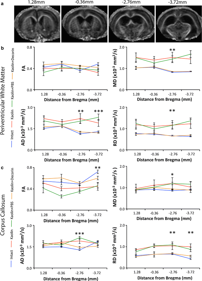
Decorin reduced hydrocephalus induced abnormalities in the caudal corpus callosum and periventricular white matter as evident from DTI. a Representative FA images of the locations at which the corpus callosum and periventricular white matter were analysed. Section 1 (1.28 mm anterior to Bregma) and Section 2 (0.36 mm posterior to Bregma) are classified as the rostral periventricular white matter and corpus callosum. Section 3 (2.76 mm posterior to Bregma) and Section 4 (3.72 mm posterior to Bregma) refer to the caudal periventricular white matter and corpus callosum. Line graphs displaying decorin’s ability to reduce abnormalities in the (b) corpus callosum and (c) periventricular white matter on DTI; blue = Intact, green = kaolin, red = kaolin + PBS, orange = kaolin + decorin [Intact (n = 4), kaolin (n = 4), kaolin + PBS (n = 6), kaolin + decorin (n = 5)]. Error bars represent the standard error of the mean; *p < 0.05, **p < 0.01, ***p < 0.001
Fig. 3
Fig. 3
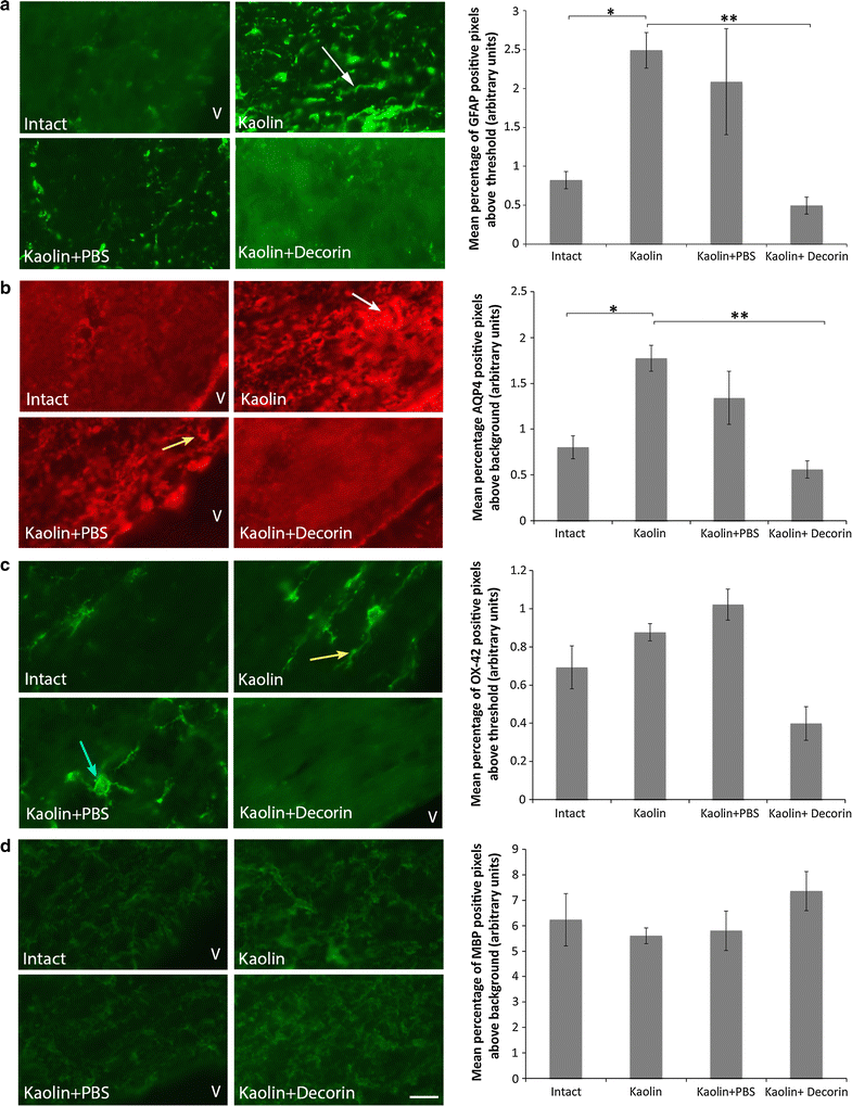
Decorin prevented an increase in GFAP and AQP4 in the periventricular white matter. Representative images comparing the level of (a) GFAP immunostaining (green), (b) AQP4 immunostaining (red), (c) OX-42 immunostaining (green) and (d) MBP immunostaining (green) in the periventricular white matter; scale bar = 10 μm. a kaolin and kaolin + PBS rats displayed thickening of astrocytic processes (white arrow). b Accumulation of AQP4 staining was observed in kaolin rats (white arrow). AQP4 was further arranged around the circumference of blood vessels (yellow arrow). c Elongated, amoeboid microglia (yellow arrow) were particularly evident in kaolin rats. Microglia of kaolin + PBS rats were captured transitioning from branched resting microglia to activated amoeboid microglia (blue arrow). d Decorin treatment improved the myelin loss and disorganisation present in kaolin and kaolin + PBS rats (white arrow). Each corresponding bar graph displays the mean percentage of GFAP, AQP4, OX-42 or MBP positive pixels above threshold or background in the periventricular white matter across the four experimental groups; V lateral ventricle, error bars represent the standard error of the mean, *p < 0.05, **p < 0.01
Fig. 4
Fig. 4
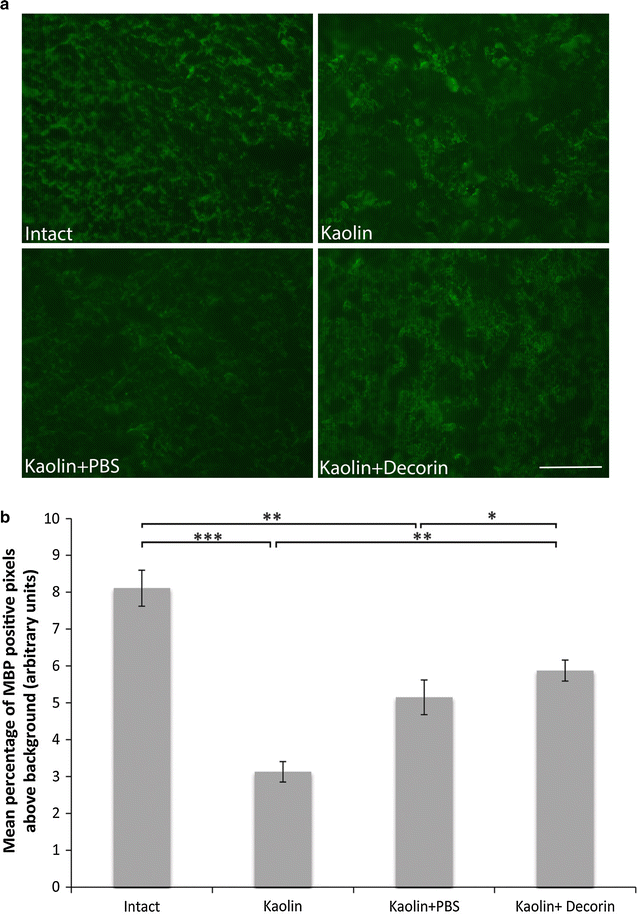
Decorin prevented myelin loss in the caudal internal capsule. a Representative images comparing caudal internal capsule MBP immunostaining (green) across the four experimental groups. Myelin organisation was better maintained with decorin use. b A bar graph displaying the mean percentage of MBP positive pixels above threshold in the internal capsule across the four experimental groups. Decreased MBP levels were present in kaolin and kaolin + PBS rats which was incompletely attenuated with decorin treatment; error bars represent the standard error of the mean, *p < 0.05, **p < 0.01, ***p < 0.001; scale bar = 50 μm

References

    1. Fernell E, Hagberg G, Hagberg B. Infantile hydrocephalus in preterm, low-birth-weight infants—a nationwide Swedish cohort study 1979–1988. Acta Pediatr. 1993;82:45–48. doi: 10.1111/j.1651-2227.1993.tb12513.x. - DOI - PubMed
    1. Simon TD, Riva-Cambrin J, Srivastava R, Bratton SL, Dean JM, Kestle JR, et al. Hospital care for children with hydrocephalus in the United States: utilization, charges, comorbidities, and deaths. J Neurosurg Pediatr. 2008;1:131–137. doi: 10.3171/PED/2008/1/2/131. - DOI - PubMed
    1. Blackburn BL, Fineman RM. Epidemiology of congenital hydrocephalus in Utah, 1940–1979: report of an iatrogenically related “epidemic”. Am J Med Genet. 1994;52:123–129. doi: 10.1002/ajmg.1320520202. - DOI - PubMed
    1. Amlashi SF, Riffaud L, Morandi X. Communicating hydrocephalus and papilloedema associated with intraspinal tumours: report of four cases and review of the mechanisms. Acta Neurol Belg. 2006;106:31–36. - PubMed
    1. Cooke RS, Patterson V. Acute obstructive hydrocephalus complicating bacterial meningitis. Hydrocephalus was probably non-obstructive. BMJ. 1999;318:124. doi: 10.1136/bmj.318.7176.124. - DOI - PubMed

MeSH terms