. 2016 Jul 29;291(31):16082-9.
doi: 10.1074/jbc.M116.734822. Epub 2016 May 31.

The CaMKII/GluN2B Protein Interaction Maintains Synaptic Strength

Affiliations

The CaMKII/GluN2B Protein Interaction Maintains Synaptic Strength

Kelsey Barcomb et al. J Biol Chem. .

Abstract

Learning, memory, and cognition are thought to require normal long-term potentiation (LTP) of synaptic strength, which in turn requires binding of the Ca(2+)/calmodulin-dependent protein kinase II (CaMKII) to the NMDA-type glutamate receptor (NMDAR) subunit GluN2B. For LTP induction, many additional required players are known. Here we tested the hypothesis that CaMKII/GluN2B binding also mediates the more elusive maintenance of synaptic strength. Intriguingly, the CaMKII inhibitor tatCN21 reduces synaptic strength only at high concentrations necessary for CaMKII/NMDAR disruption (20 μm) but not at lower concentrations sufficient for kinase inhibition (5 μm). However, increased concentration also causes unrelated effects. Thus, to distinguish between correlation and causality, we used a pharmacogenetic approach. In a mouse with a mutant NMDAR GluN2B subunit that is CaMKII binding-incompetent, any tatCN21 effects that are specific to the CaMKII/GluN2B interaction should be abolished, and any remaining tatCN21 effects have to be nonspecific (i.e. mediated by other targets). The results showed that the persistent reduction of synaptic strength by transient application of 20 μm tatCN21 had a nonspecific presynaptic component (on fiber volley amplitude) that was unrelated to the CaMKII/GluN2B interaction or CaMKII activity. However, the remaining component of the persistent tatCN21 effect was almost completely abolished in the GluN2B mutant mouse. These results highlight the requirement for stringent pharmacogenetic approaches to separate specific on-target effects from nonspecific off-target effects. Importantly, they also demonstrate that the CaMKII/GluN2B interaction is required not only for normal LTP induction but also for the maintenance of synaptic strength.

Keywords: Ca2+/calmodulin-dependent protein kinase II (CaMKII); N-methyl-d-aspartate receptor (NMDA receptor, NMDAR); glutamate receptor; hippocampus; synapse.


Figures

FIGURE 1.
FIGURE 1.
Reduction of synaptic strength by 20 μm tatCN21. A, application of 20 μm tatCN21 for 30 min after at least 20 min of stable baseline reduced basal synaptic strength, which partially recovered during a 60-min washout period in hippocampal recordings from WT slices (n = 8). 1, 2, and 3 indicate the time points shown in B. B, example fEPSP traces for one experiment from the time course in A. After baseline recording (1), tatCN21 treatment (2) reduced the response size, which partially recovered after washout (3). Merged traces are shown below, with the washout response as dotted line. C, after at least 20 min of stable baseline, LTP was induced by a four-tetanus protocol (4T; 4 × 100 Hz, 1 s) followed by an additional tetanus (1T) 20 min later to show saturation. 30 min after the initial four tetani, 20 μm tatCN21 was applied, which reduced potentiated transmission to below the initial baseline value. A 60-min washout led to a partial recovery of the response. Saturation of LTP was reversed, as an additional four tetani after washout reinduced LTP. D, the lower concentration of 5 μm tatCN21 (sufficient to block enzymatic activity) was applied after stable baseline and did not reduce basal transmission (n = 4), consistent with previous results.
FIGURE 2.
FIGURE 2.
20 μm tatCN21 differentially affects basal transmission of WT and KI mice. A, 20 μm tatCN21 was applied for 30 min after at least 20 min of stable baseline, followed by 60 min of washout in hippocampal recordings. Responses from WT slices (black circles, same as in Fig. 1) and from KI slices (green triangles) are compared. Reduction of the tatCN21 effect in the KI slice indicates a specific effect by CaMKII/GluN2B disruption in the WT slices; however, the remaining effect must be nonspecific. Results expected based on purely specific or purely nonspecific effects are indicated by dashed green lines. 1, 2, and 3 indicate the time points shown in B. B, example traces from KI slices at the time points indicated in A, representing baseline (1), drug treatment (2), and washout (3). The traces are shown overlapping below. C, quantification of the time course for baseline (−10 to −2 min), drug treatment (22–30 min, and washout (82–90 min) for both WT (n = 8) and KI (n = 6) recordings. Compared with WT slices, the responses in KI slices were significantly less reduced during both treatment and after washout (two-way ANOVA; *, p < 0.05; ***, p < 0.001). For the WT, the responses partially recovered after washout (one-way ANOVA; +, p < 0.05). For KI, the responses during treatment and after washout did not differ significantly (one-way ANOVA; NS, non-significant). In both WT and KI slices, all responses after drug application were significantly reduced compared with baseline (one-way ANOVA; p < 0.001).
FIGURE 3.
FIGURE 3.
20 μm tatCN21 persistently reduces presynaptic function. A, FV amplitudes (Amp) were measured over the same time course as in Fig. 2A. In both WT and KI slices, 20 μm tatCN21 reduced FV amplitudes to about 70%, which remained persistently reduced after washout. B, quantification of the time course for baseline (−10 to −2 min), drug treatment (22–30 min), and washout (82–90 min). FV amplitudes were significantly reduced compared with baseline during both treatment and washout for both genotypes (two-way ANOVA; *, p < 0.05). No differences were seen between WT and KI. NS, non-significant. C, no reduction in FV amplitudes was observed with 5 μm tatCN21 (n = 4), measured over the time course shown in Fig. 1D, consistent with previous results that found no change in fEPSC amplitude. D, paired pulse ratios were increased during 20 μm tatCN21 treatment in WT mice (compared with baseline; one-way ANOVA; *, p < 0.05; n = 4) and returned to baseline during washout.
FIGURE 4.
FIGURE 4.
Persistent postsynaptic reduction is nearly absent in GluN2BΔCaMKII KI slices. A, I/O curves for WT slices, generated before and after each treatment with 20 μm tatCN21. The fEPSP slopes are plotted as a function of FV amplitudes. A linear best fit line is shown to compare the I/O relationships, indicating a reduction in postsynaptic function after 20 μm tatCN21 treatment and washout (n = 4). B, I/O curves as in A for GluN2BΔCaMKII mice, revealing a smaller reduction in the I/O relationship by tatCN21 treatment (n = 3). C, comparing slopes of the best fit lines in A and B, both normalized to the baseline before tatCN21 treatment. Both WT and KI slices had a significant reduction in slope after treatment, but this reduction was much less in the KI compared with WT mice (two-way ANOVA; **, p < 0.01; ***, p < 0.001). D, calculation of the non-FV contribution to the total effect of 20 μm tatCN21 in WT slices, which approximates the postsynaptic component. Both fEPSP slope data (gray squares) and FV amplitude data (open triangles) were normalized to percent of baseline. Then the approximate postsynaptic component (Post, black circles) was calculated by subtracting the normalized FV amplitudes from the normalized fEPSP slopes. The persistent postsynaptic contribution then shows a reduction to about 55% of baseline during the persistent phase in WT slices. E, calculation of the non-FV contribution as in D but for KI mice. The persistent postsynaptic contribution for KI slices shows only a small reduction during the persistent phase.
FIGURE 5.
FIGURE 5.
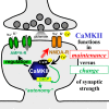
Schematic of an excitatory spine synapse. CaMKII functions in plasticity versus maintenance of synaptic strength are indicated.

References

    1. Coultrap S. J., Freund R. K., O'Leary H., Sanderson J. L., Roche K. W., Dell'Acqua M. L., and Bayer K. U. (2014) Autonomous CaMKII mediates both LTP and LTD using a mechanism for differential substrate site selection. Cell Rep. 6, 431–437 - PMC - PubMed
    1. Coultrap S. J., and Bayer K. U. (2012) CaMKII regulation in information processing and storage. Trends Neurosci. 35, 607–618 - PMC - PubMed
    1. Lisman J., Yasuda R., and Raghavachari S. (2012) Mechanisms of CaMKII action in long-term potentiation. Nat. Rev. Neurosci. 13, 169–182 - PMC - PubMed
    1. Hell J. W. (2014) CaMKII: claiming center stage in postsynaptic function and organization. Neuron 81, 249–265 - PMC - PubMed
    1. Shonesy B. C., Jalan-Sakrikar N., Cavener V. S., and Colbran R. J. (2014) CaMKII: a molecular substrate for synaptic plasticity and memory. Prog. Mol. Biol. Transl. Sci. 122, 61–87 - PubMed

Publication types

MeSH terms

Substances