Review
. 2016 Dec;18(12):1758-1772.
doi: 10.1093/europace/euw014. Epub 2016 May 31.

The role of myocardial wall thickness in atrial arrhythmogenesis

Affiliations
Review

The role of myocardial wall thickness in atrial arrhythmogenesis

John Whitaker et al. Europace. 2016 Dec.

Abstract

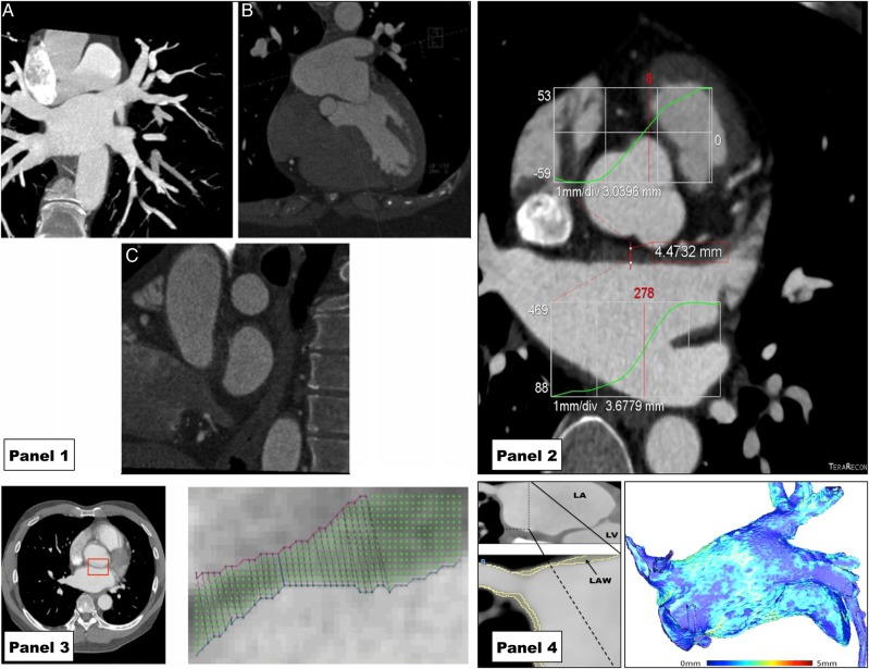
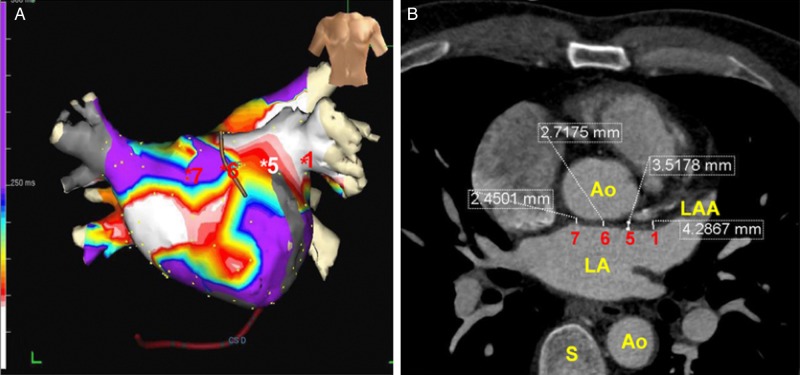
Changes in the structure and electrical behaviour of the left atrium are known to occur with conditions that predispose to atrial fibrillation (AF) and in response to prolonged periods of AF. We review the evidence that changes in myocardial thickness in the left atrium are an important part of this pathological remodelling process. Autopsy studies have demonstrated changes in the thickness of the atrial wall between patients with different clinical histories. Comparison of the reported tissue dimensions from pathological studies provides an indication of normal ranges for atrial wall thickness. Imaging studies, most commonly done using cardiac computed tomography, have demonstrated that these changes may be identified non-invasively. Experimental evidence using isolated tissue preparations, animal models of AF, and computer simulations proves that the three-dimensional tissue structure will be an important determinant of the electrical behaviour of atrial tissue. Accurately identifying the thickness of the atrial may have an important role in the non-invasive assessment of atrial structure. In combination with atrial tissue characterization, a comprehensive assessment of the atrial dimensions may allow prediction of atrial electrophysiological behaviour and in the future, guide radiofrequency delivery in regions based on their tissue thickness.

Keywords: AF; Atrial fibrillation; Cardiac CT; Cardiac computed tomography; Left atrial wall thickness.


Figures

Figure 1
Figure 1
Human atrial anatomy: pathological specimens with corresponding anatomical landmarks from Carto electro-anatomical map of right atrium under anteroposterior projection (purple) and left atrium under modified left anterior oblique projection (green). ( A–E) Reproduced with permission of BMJ publishing group from Wang et al. (A) Right atrium opened through an incision that passes parallel to the right coronary artery to the tip of the appendage and then through the terminal crest (O–O) into the superior vena cava. Pectinate muscles arise along the length of the terminal crest (+ +) towards the coronary sinus (*). There is a prominent Eustachian valve in this specimen. (B) Atria viewed from behind showing intercaval bundle after pericardium removed. (C) Tricuspid valve viewed from the right atrium with the endocardium removed to reveal the internal circumferential bundle (arrows) encircling the vestibule. The pectinate muscles (lines) are perpendicular to the circumferential bundle. ‘+’ indicates terminal crest. (D) External view of left atrium showing tubular appendage with arrow junction to pulmonary veins. (E) Internal aspect of left atrium demonstrating fossa ovalis on the septum. Pectinate muscles are confined to the appendage. The vestibule leading to the mitral valve (MV) is smooth.
Figure 2
Figure 2
Tissue section of human atria reproduced with permission from Cabrera et al. (A) Endocardial surface of the left lateral wall and (B) with translumination showing the prominent lateral ridge(*) interposed between the left atrial appendage (LAA) and the left pulmonary veins. Muscular trabeculations denoted by red arrows extend inferiorly from the ridge to the vestibule. The dotted line marks the mitral valve (MV) annulus. Lines ‘a’ and ‘b’ mark the sites where measurements were taken in this study. (C) is a section through line ‘a’ to demonstrate the thickness of the ridge. Also seen in the left circumflex coronary artery (LCX), coronary sinus (CS), and left ventricle (LV).
Figure 3
Figure 3
Recent examples of the use of cardiac CT to assess LAWT demonstrating evolving methodology. Panel 1 reproduced with permission from Beinart et al. Standardized planes for measuring LAWT manually. (A) Oblique coronal plane parallel to posterior wall of LA (in maximum intensity projection). (B) Axial plane perpendicular to coronal plane at the level of the mitral isthmus. (C) Saggital plane orthogonal to the original plane near the ostium of the left pulmonary vein. Panel 2 reproduced with permission from Wi et al. Semi-automatic measurement of the LAWT based on histogram intensities (projected onto CT image) after manually selecting a region containing the LA wall. Panel 3 reproduced with permission from Dewland et al. An example of an automated, computer-based segmentation of the LA wall. (A) Axial plane of contrast-enhanced cardiac CT with anterior LA wall identified by red box. (B) An expanded illustration of this region with LA wall identified by computer algorithm on the basis of voxel intensity. Panel 4: a semi-automated approach to LA wall segmentation based on HU intensity that has been used to generate whole chamber LAWT maps as reported by Bishop et al. Left hand image demonstrates a multiplanar-reconstructed CT image of the left atrium. Expanded region shows the LA roof and right superior pulmonary vein with LA wall as identified by computer algorithm in yellow. Right image is a visual representation of the LAWT throughout the chamber calculated using the method reported by Bishop et al. based on generating field lines and solving the Laplace equation.
Figure 4
Figure 4
The correlation of CT-measured LAWT with clinically measurable electrophysiological phenomena. Reproduced with permission from Wi et al. Left hand panel (A) is colour map representing the summation of CFAEs across the left atrium under an anteroposterior projection. On the left hand side (B) is the CT image from which LAWT measurements were made with representative measurements. In this report, CFAEs were observed more frequently in regions with increased atrial wall thickness (sites 1 and 5).

References

    1. Becker AE. Left atrial isthmus: anatomic aspects relevant for linear catheter ablation procedures in humans. J Cardiovasc Electrophysiol 2004;15:809–12. - PubMed
    1. Hall B, Jeevanantham V, Simon R, Filippone J, Vorobiof G, Daubert J. Variation in left atrial transmural wall thickness at sites commonly targeted for ablation of atrial fibrillation. J Interv Card Electrophysiol 2006;17:127–32. - PubMed
    1. Sánchez-Quintana D, Cabrera JA, Climent V, Farré J, De Mendonça MC, Ho SY. Anatomic relations between the esophagus and left atrium and relevance for ablation of atrial fibrillation. Circulation 2005;112:1400–5. - PubMed
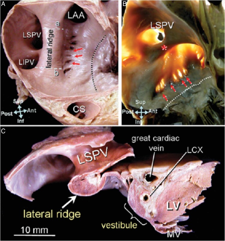
    1. Cabrera JA, Ho SY, Climent V, Sánchez-Quintana D. The architecture of the left lateral atrial wall: a particular anatomic region with implications for ablation of atrial fibrillation. Eur Heart J 2008;29:356–62. - PubMed
    1. Platonov PG, Ivanov V, Ho SY, Mitrofanova L. Left atrial posterior wall thickness in patients with and without atrial fibrillation: data from 298 consecutive autopsies. J Cardiovasc Electrophysiol 2008;19:689–92. - PubMed