Review
. 2016 Aug:44:123-135.
doi: 10.1016/j.dnarep.2016.05.018. Epub 2016 May 20.

XPA: A key scaffold for human nucleotide excision repair

Affiliations
Review

XPA: A key scaffold for human nucleotide excision repair

Norie Sugitani et al. DNA Repair (Amst). 2016 Aug.

Abstract

Nucleotide excision repair (NER) is essential for removing many types of DNA lesions from the genome, yet the mechanisms of NER in humans remain poorly understood. This review summarizes our current understanding of the structure, biochemistry, interaction partners, mechanisms, and disease-associated mutations of one of the critical NER proteins, XPA.

Keywords: DNA repair; NER; XPA; Xeroderma pigmentosum..


Figures

Figure 1
Figure 1. Domain map of XPA and interaction partners
Schematic domain structure of human XPA protein (top). The region containing the globular core is colored pink, with the location of the Zn finger indicated as a yellow circle. The nuclear localization signal (NLS) is colored yellow. The N- and C-termini are dynamically disordered. Known interaction partners are shown below the domain map, aligned with the XPA residues involved in each interaction. Gray proteins are those known to interact with XPA but for which the sites of interaction have not been determined. Blue indicates a binding partner for which the binding sites on XPA remain controversial. If known, the domain or residues involved in XPA binding are given in parenthesis.
Figure 2
Figure 2. A structure of the globular core of XPA
Left - surface representation of the solution NMR structure of the globular core of XPA (PDB: 1XPA) colored by electrostatic field at the surface. Positive charge is in blue tones and negative charge in red tones. Right – Ribbon diagram of 1XPA.
Figure 3
Figure 3. Structures of S. cerevisiae Rad14 in complex with DNA
A) Upper panel, x-ray crystal structure of Rad14t (dark and light green) bound to a cis-platin-containing duplex (PDB: 5A39). Lower panel, sequence of the DNA duplex. B) Upper panel, x-ray crystal structure of Rad14t molecules (purple and pink) bound to an AAF-containing duplex (PDB: 5A3D). Lower panel, sequence of the DNA duplex.
Figure 4
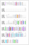
Figure 4. Alignment of the XPA protein sequence across seven diverse species
A structure-guided sequence alignment of XPA proteins from seven species. The extent and secondary structure in human XPA98-219 construct as determined in the NMR structure (PDB: 1XPA) is indicated above the alignment. The residues not visible in the structure are indicated with the dotted line above the sequence. The secondary structure in the S. cerevisiae Rad14188-302 construct as determined in the crystal structures (PDB: 5A39, 5A3D) is given below the alignment. The 20-residue extension of XPA required for full DNA binding is also highlighted. Asterisks mark residues identified as critical for DNA binding in the Rad14 crystal structures. The alignment was computed by PROMALS3D using 1XPA_A and 5A3D_A as guides. Residues are colored and conserved alignment columns are boxed according to the default similarity scores in ESPript.
Figure 5
Figure 5. Evolutionary conservation of XPA
The surface representation of the globular core of human XPA (PDB: 1XPA) colored by evolutionary conservation computed from the alignment of orthologous XPA sequences from human, mouse, chicken, frog, fruit fly, fission yeast, and baker’s yeast (Figure 4). The rendering of the structure was created with Chimera.
Figure 6
Figure 6. Comparing structures of human XPA with S. cerevisiae Rad14
A) One molecule from the crystal structure of Rad14t bound to a cis-platin-containing duplex (PDB: 5A3D). B) A representative conformer from the NMR solution structure of the globular core of human XPA (PDB ID:1XPA). C) Overlay of structures in panels A and B.
Figure 7
Figure 7. Structures of XPA in complex with other NER proteins
A) Schematic domain map of human RPA. DNA binding domains (A, B, C, D) have stipled shading. Domains involved in protein interactions are underlined, with those involved in XPA interactions in pink. B) Ribbon diagram of the solution NMR structure (PDB: 1DPU) of RPA32C (light green) in complex with a peptide fragment of UNG2 (salmon), which binds to RPA32C in the same manner as XPA29-46. C) X-ray crystal structure of a peptide fragment of XPA (salmon) in complex with ERCC1 (violet) (PDB: 2JNW). XPA residue numbers are indicated in panels B and C.
Figure 8
Figure 8. Model of some XPA interactions in NER incision complexes
An homology model of XPA102-214 in complex with an AAF-containing duplex was built based on the Rad14t structure (PDB: 5A3D). A SAXS model was used for the RPA DNA binding core in complex with ssDNA. The structure of RPA70N is taken from an X-ray crystal structure (PDB: PDB: 1EWI structure). The structure of RPA32C in complex with a peptide fragment of UNG2 (PDB: 1DPU) was used to represent RPA32C bound to XPA29-46. The XPF-ERCC1 model combined ERCC196-214 in complex with XPA67-80, XPF842-916 in complex with ssDNA (PDB: 2KN7), and ERCC1220-297 (PDB: 1Z00). Dashed lines indicate potential path of linkers or DNA. The DNA lesion is represented by a red star. Colors: XPA – pink, RPA70 – blue, RPA32 – green, RPA14 – dark red, XPF – purple, ERCC1 – violet, DNA – dark grey.
Figure 9
Figure 9. XPA gene structure and mutations
The blue boxes give a schematic representation of the human XPA gene structure. Exons are represented by large boxes, introns by medium boxes, and introns by blue lines connecting the exons. Colored ellipses within the gene model show the location and frequency of XPA mutations observed in the ExAC database of 60,706 human exome sequences. Missense mutations and inframe indels are colored yellow; frameshifts, gained stop codons, and mutations to splice acceptor/donor sites are colored red; synonymous mutations are in green; and non-coding variants are colored black. The eccentricity of each ellipse indicates the mutation’s frequency in the ExAC population. Coding variation is rare in XPA; the most common coding variant has a frequency of 0.3%.

References

    1. Scharer OD. Nucleotide Excision Repair in Eukaryotes. Cold Spring Harb. Perspect. Biol. 2013;5:1–19. - PMC - PubMed
    1. Gillet LCJ, Scharer OD. Molecular Mechanisms of Mammalian Global Genome Nucleotide Excision Repair. Chem. Rev. 2006;106:253–276. - PubMed
    1. Truglio JJ, Croteau DL, Houten B. Van, Kisker C. Prokaryotic Nucleotide Excision Repair : The UvrABC System. 2006. - PubMed
    1. DiGiovanna JJ, Kraemer KH. Shining a light on xeroderma pigmentosum. J. Invest. Dermatol. 2012;132:785–96. - PMC - PubMed
    1. Hoeijmakers JHJ. DNA damage, aging, and cancer. N. Engl. J. Med. 2009;361:1475–85. - PubMed

Publication types

MeSH terms