. 2016 Aug 12;36(17):2226-35.
doi: 10.1128/MCB.01102-15. Print 2016 Sep 1.

The C-Terminal RGG Domain of Human Lsm4 Promotes Processing Body Formation Stimulated by Arginine Dimethylation

Affiliations

The C-Terminal RGG Domain of Human Lsm4 Promotes Processing Body Formation Stimulated by Arginine Dimethylation

Marcos Arribas-Layton et al. Mol Cell Biol. .

Abstract

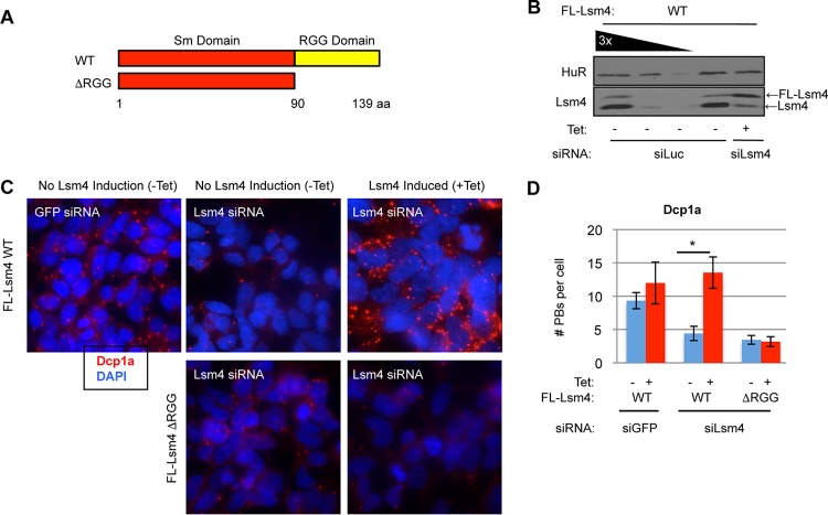
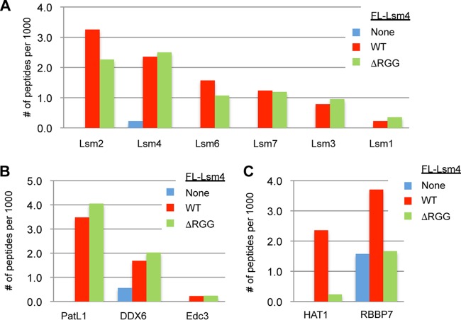
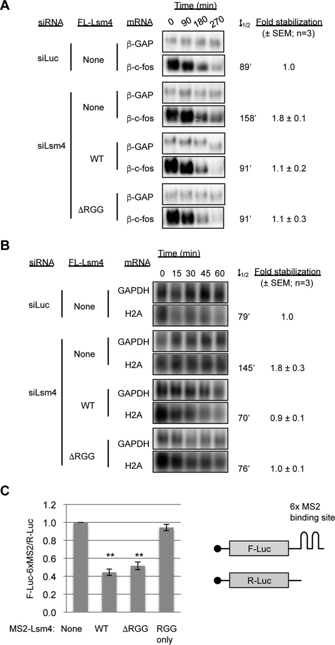
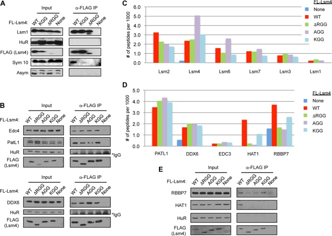
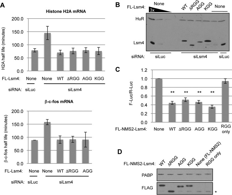
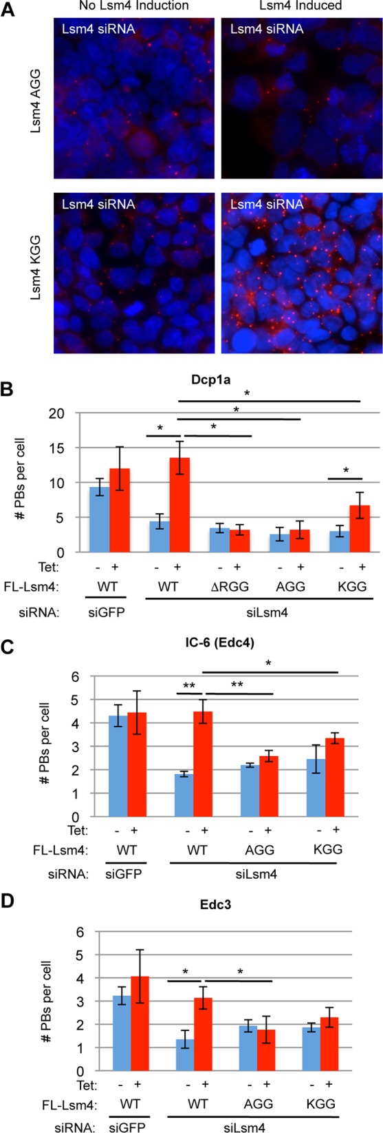
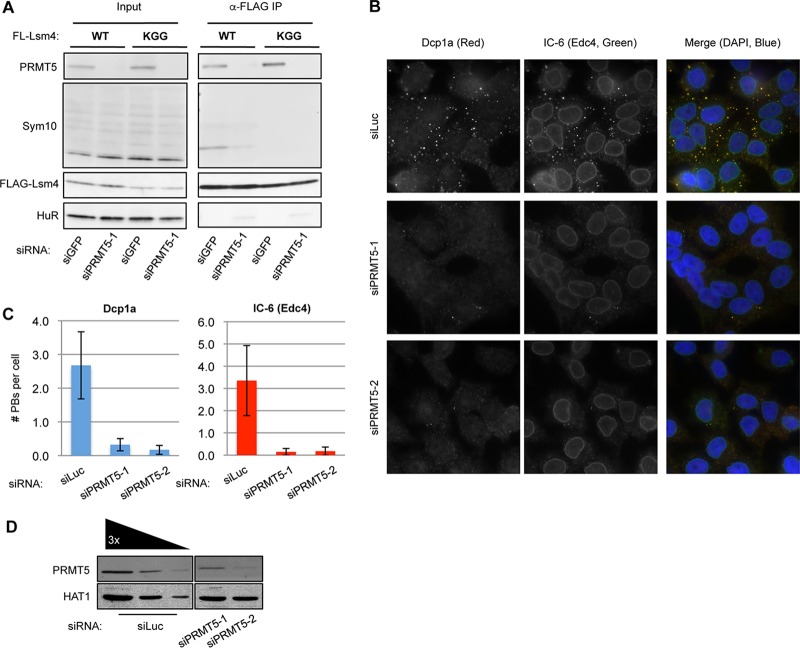
Processing bodies (PBs) are conserved cytoplasmic aggregations of translationally repressed mRNAs assembled with mRNA decay factors. The aggregation of mRNA-protein (mRNP) complexes into PBs involves interactions between low-complexity regions of protein components of the mRNPs. In Saccharomyces cerevisiae, the carboxy (C)-terminal Q/N-rich domain of the Lsm4 subunit of the Lsm1-7 complex plays an important role in PB formation, but the C-terminal domain of Lsm4 in most eukaryotes is an RGG domain rather than Q/N rich. Here we show that the Lsm4 RGG domain promotes PB accumulation in human cells and that symmetric dimethylation of arginines within the RGG domain stimulates this process. A mutant Lsm4 protein lacking the RGG domain failed to rescue PB formation in cells depleted of endogenous Lsm4, although this mutant protein retained the ability to assemble with Lsm1-7, associate with decapping factors, and promote mRNA decay and translational repression. Mutation of the symmetrically dimethylated arginines within the RGG domain impaired the ability of Lsm4 to promote PB accumulation. Depletion of PRMT5, the primary protein arginine methyltransferase responsible for symmetric arginine dimethylation, including Lsm4, resulted in loss of PBs. We also uncovered the histone acetyltransferase 1 (HAT1)-RBBP7 lysine acetylase complex as an interaction partner of the Lsm4 RGG domain but found no evidence of a role for this complex in PB metabolism. Together, our findings suggest a stimulatory role for posttranslational modifications in PB accumulation and raise the possibility that mRNP dynamics are posttranslationally regulated.


Figures

FIG 1
FIG 1
The RGG domain of Lsm4 is required for visible PBs. (A) Schematic showing WT and ΔRGG mutant Lsm4 proteins. aa, amino acids. (B) WB assay showing depletion of endogenous Lsm4 and expression of exogenous WT Lsm4 at nearly endogenous levels in stable HEK 293 T-REx cells. The first three lanes (from the left) are loaded with 100, 33, and 11% of the total protein in the last two lanes. (C) HEK 293 T-REx cells treated with siRNA against GFP (control) or Lsm4, induced to express FLAG (FL)-tagged WT or ΔRGG mutant Lsm4, and stained for Dcp1a (red). DAPI staining is blue. (D) Quantification of PB numbers from three independent indirect IF experiments. Error bars represent standard deviations. *, P < 0.05 (Student's paired two-tailed t test).
FIG 2
FIG 2
The RGG domain of Lsm4 is required for interaction with HAT1/RBBP7 but is not necessary for association with Lsm1-7 components or decapping factors. The graphs show the numbers of peptides detected per 1,000 total peptides coprecipitating with FLAG-tagged WT or ΔRGG mutant Lsm4 for Lsm1-7 complex members (A), decapping factors (B), or HAT1 complex members (C). None is a negative-control FLAG (FL) IP performed with parental cells containing no FLAG-Lsm4 protein. See Table S1 in the supplemental material for the primary data.
FIG 3
FIG 3
The RGG domain of Lsm4 is not required for mRNA decay or translational repression. (A) Northern blot assays showing the decay of an ARE-containing reporter mRNA (β-c-fos) in HeLa Tet-off cells treated with the siRNAs indicated and transiently expressing the Lsm4 proteins indicated. β-c-fos half-lives (t1/2) were calculated with the constitutively transcribed β-GAP internal control mRNA for normalization. Fold stabilization was calculated relative to the siLuc control condition in three experiments, and the standard errors of the means are indicated. (B) Northern blot assays showing the decay of endogenous H2A mRNA induced by treatment with 5 mM hydroxyurea in HEK 293 T-REx cells treated with the siRNAs indicated and stably expressing the Lsm4 proteins indicated. Glyceraldehyde 3-phosphate dehydrogenase (GAPDH) mRNA served as a normalization control. (C) Luciferase luminescence assays of cells transiently transfected with plasmids encoding the MS2 fusion proteins indicated, as well as a firefly luciferase reporter with six MS2 coat protein binding sites in its 3′ UTR (F-Luc-6xMS2) and a Renilla luciferase reporter (R-Luc) as an internal control. F-Luc-6xMS2 was normalized to R-Luc, and all samples were normalized to cells transfected with MS2 and the reporters alone. Error bars represent the standard errors of the means. **, P < 0.01 (Student's paired two-tailed t test).
FIG 4
FIG 4
Arginines in the Lsm4 RGG domain are required for interaction with Hat1/RBBP7 but dispensable for association with Lsm1-7 and decapping factors. (A, B) WB assay showing Lsm1 and decapping factors coimmunoprecipitating with WT and mutant Lsm4. Input samples are shown on the left. Sym10 is specific for symmetrically dimethylated arginines, and Asym is specific for asymmetrically dimethylated arginines. The asterisk indicates cross-reaction with IgG. (C, D) Graphs showing the numbers of peptides detected per 1,000 total peptides from Lsm factors, decapping factors, and Hat1 complex members coprecipitating with WT or mutant Lsm4, as indicated. (E) WB assay showing HAT1 complex members coimmunoprecipitating with WT and mutant Lsm4.
FIG 5
FIG 5
Arginines in the Lsm4 RGG domain are dispensable for mRNA repression and degradation. (A) Graphs showing histone H2A and β-c-fos mRNA half-lives in the presence of WT and mutant Lsm4 monitored as described in the legend to Fig. 3A and B. Error bars represent the standard errors of the means. (B) WB assay showing nearly endogenous expression of FL-Lsm4 proteins in the stable cell lines used in panel A. (C) Graphs showing luciferase activity from tethering of MS2-Lsm4 WT and mutant proteins monitored as described in the legend to Fig. 3C. Error bars represent the standard errors of the means. **, P < 0.01 (Student's paired two-tailed t test). (D) WB assay showing even expression of FL-MS2-Lsm4 proteins as detected by the FLAG antibody. The asterisk indicates a cross-reacting band.
FIG 6
FIG 6
Arginines in the Lsm4 RGG domain are required for PB accumulation. (A) Indirect IF assays for Dcp1a (red) in HEK 293 T-REx cells treated with an siRNA against Lsm4 and induced to express the AGG and KGG mutant Lsm4 proteins. DAPI staining is shown in blue to mark nuclei. (B to D) Quantification of PBs by the PB markers indicated in three independent experiments. Error bars represent standard deviations. **, P < 0.01; *, P < 0.05 (Student's paired two-tailed t test).
FIG 7
FIG 7
PRMT5 is required for PB formation. (A) WB assay monitoring symmetric arginine dimethylation (with Sym10) in FLAG-tagged WT Lsm4 and KGG immunoisolated from cells treated with siGFP and siPRMT5-1 siRNAs. (B) Indirect IF assays for Dcp1a (red) and Edc4 (IC-6; green) in HeLa cells treated with an siRNA against luciferase (control) or PRMT5 (two different siRNAs). DAPI staining is shown in blue to mark nuclei. Merged images are on the right. (C) Quantification of PBs for Dcp1a and Edc4. Error bars represent standard deviations. (D) WB assay showing PRMT5 depletion with HAT1 serving as a loading control. The first three lanes (from the left) are loaded with 100, 33, and 11% of the total protein in the last two lanes.

References

    1. Guhaniyogi J, Brewer G. 2001. Regulation of mRNA stability in mammalian cells. Gene 265:11–23 . doi:10.1016/S0378-1119(01)00350-X. - DOI - PubMed
    1. Parker R, Sheth U. 2007. P bodies and the control of mRNA translation and degradation. Mol Cell 25:635–646. doi:10.1016/j.molcel.2007.02.011. - DOI - PubMed
    1. Wells SE, Hillner PE, Vale RD, Sachs AB. 1998. Circularization of mRNA by eukaryotic translation initiation factors. Mol Cell 2:135–140. doi:10.1016/S1097-2765(00)80122-7. - DOI - PubMed
    1. Chen C-YA, Shyu A-B. 2011. Mechanisms of deadenylation-dependent decay. Wiley Interdiscip Rev RNA 2:167–183. doi:10.1002/wrna.40. - DOI - PMC - PubMed
    1. Huch S, Nissan T. 2014. Interrelations between translation and general mRNA degradation in yeast. Wiley Interdiscip Rev RNA 5:747–763. doi:10.1002/wrna.1244. - DOI - PMC - PubMed

Publication types

MeSH terms