. 2016 Jun 14;113(24):E3413-22.
doi: 10.1073/pnas.1606636113. Epub 2016 May 31.

Envelope residue 375 substitutions in simian-human immunodeficiency viruses enhance CD4 binding and replication in rhesus macaques

Affiliations

Envelope residue 375 substitutions in simian-human immunodeficiency viruses enhance CD4 binding and replication in rhesus macaques

Hui Li et al. Proc Natl Acad Sci U S A. .

Abstract

Most simian-human immunodeficiency viruses (SHIVs) bearing envelope (Env) glycoproteins from primary HIV-1 strains fail to infect rhesus macaques (RMs). We hypothesized that inefficient Env binding to rhesus CD4 (rhCD4) limits virus entry and replication and could be enhanced by substituting naturally occurring simian immunodeficiency virus Env residues at position 375, which resides at a critical location in the CD4-binding pocket and is under strong positive evolutionary pressure across the broad spectrum of primate lentiviruses. SHIVs containing primary or transmitted/founder HIV-1 subtype A, B, C, or D Envs with genotypic variants at residue 375 were constructed and analyzed in vitro and in vivo. Bulky hydrophobic or basic amino acids substituted for serine-375 enhanced Env affinity for rhCD4, virus entry into cells bearing rhCD4, and virus replication in primary rhCD4 T cells without appreciably affecting antigenicity or antibody-mediated neutralization sensitivity. Twenty-four RMs inoculated with subtype A, B, C, or D SHIVs all became productively infected with different Env375 variants-S, M, Y, H, W, or F-that were differentially selected in different Env backbones. Notably, SHIVs replicated persistently at titers comparable to HIV-1 in humans and elicited autologous neutralizing antibody responses typical of HIV-1. Seven animals succumbed to AIDS. These findings identify Env-rhCD4 binding as a critical determinant for productive SHIV infection in RMs and validate a novel and generalizable strategy for constructing SHIVs with Env glycoproteins of interest, including those that in humans elicit broadly neutralizing antibodies or bind particular Ig germ-line B-cell receptors.

Keywords: CD4; HIV-1 envelope; HIV/AIDS; SHIV.


Conflict of interest statement

The authors declare no conflict of interest.

Figures
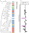
Fig. S1.
Fig. S1.
Evolutionary selection at Env residue 375 across the phylogenetic spectrum of human and simian immunodeficiency viruses. (A) A Pol (862 aa) tree of sequences from the Los Alamos HIV Sequence Database (www.hiv.lanl.gov/content/sequence/HIV/mainpage.html) inferred by using PhyML (v3.0) and an RtREV substitution model. (Scale bar: 5% genetic distance of corrected substitutions per site.) Numbers at nodes in the phylogram indicate bootstrap support ≥70% among 100 replicates. The frequency of different Env375 amino acids is represented proportionally in boxes to the right of the phylogram. Env375S is the most prevalent variant, whereas Env375H is largely restricted to the circulating recombinant form CRF01_AE. (B) Differences in average amino acid volume (117) between HIV-1 group M (3551 sequences excluding circulating recombinant forms) and SIV (292 sequences including HIV-2 and HIV-1 groups N, O, and P) for Env residues proximal (within 7 Å) to CD4 based on PDB ID code 2NXZ (85) are depicted. The two largest differences, at residues 375 and 458, are highlighted in magenta.
Fig. S2.
Fig. S2.
Sequence and structural comparisons of human and rhesus CD4 in their interactions with HIV-1 Env gp120. (A) Sequence alignment of human and rhesus CD4, with blue indicating interactions with HIV-1 Env (filled circles, both main chain and side chain; open circles, main chain only; sunbursts, side chain only). We note that the T17N substitution introduces a glycosylation sequon into the rhesus CD4. (B) Closed-prefusion trimer (PDB ID code 4TVP) with one CD4 aligned to the binding site. gp120 is shown in gray with gp41 shown in black; CD4 is shown in yellow with sequence differences between human and rhesus highlighted in green. (C) Overlay of a prefusion closed protomer (gray) with a CD4-bound gp120 (blue) and select residues highlighted. (D) Atomic details of CD4-binding in the prefusion closed context. CD4 was aligned to the prefusion closed structure using gp120. Phe-43 of CD4 is shown in stick representation along with a salt bridge formed by Asp-368 of gp120 and residue 59 of CD4, which is an Arg in human and Lys in rhesus. (E) Corresponding atomic details of the CD4-bound gp120 conformation. (F) Prefusion closed details showing the natural cavity in surface representation using the native serine residue. Mutations are shown with a stick representation of residues Ser, His, and Met shown to fill the cavity. (G) N39I makes no direct contacts with gp120 but is close to Phe-43 and could fold into the hydrophobic pocket, changing the angle of the Phe43 binding loop. (H) Overlap clash scores calculated by Molprobity for mutated residues in each conformation (using PDB ID codes 4TVP and 5CEZ for the closed form). Crystal structures of CD4-bound gp120 have previously been solved for 375S, 375H, and 375W (bold) [PDB ID codes 1RZJ (101), 3NGB (116), and 2NXZ (85)]. Rotamers with the lowest overlap were selected, although no adjustments were made to optimize surrounding side chains when calculating overlap.
Fig. 1.
Fig. 1.
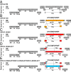
SHIV construction scheme. (AC) A complete tatrevvpuenv(gp160) segment of HIV-1 B.YU2 or D.191859 was substituted for the corresponding region in SIVmac766. (D and E) SHIV.D.191859.dCT, which lost cloning site linkers (yellow bars in C) and the carboxy terminal 33 amino acids of HIV-1 gp41 but with the carboxy terminus of SIVmac766 gp41 joined in-frame (Figs. S3 and S5A), served as a platform for further SHIV constructions containing vpuenvgp140 segments of HIV-1 strains CH505, CH848, and BG505. Met-M, Tyr-Y, His-H, Trp-W, and Phe-F were exchanged for the wild-type Ser-S residue at position 375 in Env. SIVmac766 and SHIV sequences are publicly available (GenBank accession nos. KU955514 and KU958484-9).
Fig. S3.
Fig. S3.
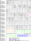
Amino acid alignment of SHIV Envs and HXB2. Contact residues with huCD4 are indicated. The SIVmac766 gp41 carboxy terminal tail exchange for the corresponding region of HIV-1 is highlighted in blue, and the HIV-1 D.191859 gp41 sequences shared with other SHIVs are highlighted in green.
Fig. 2.
Fig. 2.
SHIV Env binding to human and rhesus CD4. (A) Inhibition of SHIV entry into TZM-bl cells by human and rhesus CD4-Ig. Controls included wild-type HIV-1 Env375S expressed from infectious molecular proviral clones or as Env-pseudotyped particles. (B) SHIV entry into TZM-bl or ZB5 cells. RLU, relative light units. Variance is expressed as SD. (C) SPR analysis of HIV-1 D.191859 gp120 Env375 variants to human and rhesus CD4-Ig. Plots are of representative experiments replicated three times.
Fig. 3.
Fig. 3.
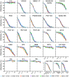
Neutralization of SHIV.BG505.332N.dCT Env375 variants in TZM-bl cells by mAbs, plasma from an HIV-1 patient with bNAbs (CH1754), HIV-1 immune globulin (HIVIG-C), or the fusion inhibitor T1249. Concentrations on the x axis are expressed in micrograms per milliliter except for CH1754, which is expressed as a plasma dilution. Plots are of representative experiments replicated three times.
Fig. S4.
Fig. S4.
Neutralization of SHIV Env375 variants in TZM-bl cells by mAbs, HIV-1 patient plasma (CH1754), HIV immune globulin (HIVIG-C), or the fusion inhibitor T1249. (A) SHIV.C.CH505. (B) SHIV.C.CH848. (C) SHIV.B.YU2. (D) SHIV.D.191859. (E) SHIV.A.BG505.332T. For SHIV.A.BG505.332T, neutralization data are shown only for the N332 glycan-dependent mAbs, because the neutralization profiles for all other mAbs were indistinguishable from SHIV.A.BG505.332N (Fig. 3). Plots are of representative experiments replicated three times.
Fig. 4.
Fig. 4.
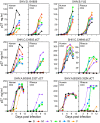
Replication of SHIV Env375 variants in primary human and rhesus CD4 T cells. Imput virus was normalized by p27Ag (SI Materials and Methods). All variants replicated efficiently in human cells, but only some variants replicated well in rhesus cells. Plots are of representative experiments replicated three times.
Fig. 5.
Fig. 5.
SHIV replication and selection in RMs. (A) CD8-depleted RMs were inoculated intravenously with a mixture of six Env375 variants (50 ng of p27Ag of each variant) of SHIV D.191859 (RM131 and RM138) or SHIV B.YU2 (RM72 and RM74). (B) Non-CD8-depleted RMs were inoculated intravenously or intrarectally with a mixture of SHIV D.191859 variants with or without a S375M substitution and with or without an SIV-for-HIV gp41 carboxy terminal tail exchange (50 ng of p27Ag of each variant intravenously; 500 ng of p27Ag of each variant intrarectally). (C and D) CD8-depleted RMs were inoculated intravenously with a mixture of six Env375 variants of SHIV C.CH505.dCT or SHIV C.CH848.dCT, respectively (50 ng of p27Ag of each variant, blue; 500 ng of p27Ag of each variant, red). (E) Non-CD8-depleted RMs inoculated intravenously with a mixture of six Env375 variants of SHIV A.BG505.dCT with or without a T332N substitution (50 ng of p27Ag of each variant; see J for inocula). (FJ) Sequence analysis of SHIV inocula and SHIV evolution in infected RMs. Sequence depth was >2,500 sequences for each MiSeq analysis (F and HJ) and ≥24 for SGS. The 293T-derived virus stock was used for all inoculations. Animal deaths are indicated by a red cross.
Fig. S5.
Fig. S5.
Identification and characterization of an SIV-for-HIV gp41 carboxy terminal tail substitution in SHIV.D.191859 infected RMs. (A) SGS of vRNA from serial plasma samples of RM131 and RM138 are shown. Strong selection leading to complete replacement of T/F sequences by sequences containing a nearly identical gp41 tail substitution is evident by 10 wk postinfection in both RMs, with partial sequence replacement as early as 4 wk. (B) Env/Gag ratios of SHIV.D.191859 with and without 375M and gp41 tail substitutions. Viruses were produced in 293T or primary human or rhesus CD4 T cells. Results are representative of two experiments. (C) Relative replication rates of SHIV.D.191859 with and without 375M and gp41 tail substitutions compared with the corresponding HIV-1 D.191859 infectious molecular clone derived virus (52). Multiplicity of infection was normalized to ∼0.3 on TZM-bl cells. Results are representative of three experiments. DPI, days postinfection.
Fig. S6.
Fig. S6.
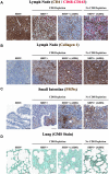
Immunopathogenesis of SHIV infections. (A and B) SHIV infection leads to progressive CD4+ T-cell loss (A) and fibrotic collagen deposition (B) in lymphoid tissues in all animals, with or without AIDS. Animals that progressed to end-stage AIDS demonstrated more severe CD4+ T-cell loss and fibrosis. (C) SHIV infection leads to GI tract damage as evidenced by infiltration of neutrophils into the lamina propria of the small and large bowel. (D) SHIV infection leads to end-stage AIDS with opportunistic infections, including PCP shown here by histopathological staining of Pneumocystis organisms in the lungs of two animals with AIDS. Animal specimens left to right: (A) RM74 preinfection, RM74 chronic infection, RM6069 and RM6447 AIDS, immunolabeled with anti-CD4, CD68–CD163. (B) RM74 preinfection, RM74 chronic infection, RM6069 and RM6447 AIDS, stained for collagen 1. (C) ZH38 (SIV/SHIV), RM74 chronic infection, RM6069 and RM6447 AIDS, stained for myeloperoxidase. (D) P376 (SIV/SHIV), RM74 chronic infection, RM6069 and RM6447 AIDS, stained with Grocott’s methenamine silver. (Scale bar: 100 μm.)
Fig. S7.
Fig. S7.
Pathological findings in SHIV infected RMs. (A) Hyperplastic Peyer’s patches with confluent germinal centers, characteristic for the asymptomatic phase of SIV infection (RM72). (B) Severe villous atrophy and crypt disruption associated with severe fibrosis of the lamina propria; numerous crypt abscesses (RM138). (C) Accumulation of abundant foamy macrophages in the lamina propria, characteristic for atypical mycobacteria opportunistic infection (RM131). (D) Lymphoid atrophy characteristic to late stages of SIV infection. Subcapsular and sinusoidal fibrosis and numerous focal hyaline deposits in the germinal centers and in the degenerated follicles. The lymph nodes are populated mainly by histiocytes, which are frequent in the enlarged lymphoid sinuses (RM74). (E) Deposition of amyloid in the germinal centers (RM74). (F) The lymphoid sinuses are dilated and filled with sheets of foamy histiocytes, a lesion characteristic for Mycobacterium avium intracellulare, an atypical mycobacterial infection commonly found in patients and RMs with AIDS (RM131). (G) Numerous lymphoid aggregates are present in the lung parenchyma and are characteristic of chronic progressive SIV infection (RM74). (H) Numerous lymphoid aggregates are present in the kidney parenchyma and are characteristic of chronic progressive SIV infection (RM138). (I) Granulomatous inflammation represented by Langerhans giant cells surrounded by numerous macrophages and lymphocytes. Numerous granulomas are present in the liver in the hepatic lobules. These lesions are characteristic of atypical mycobacterial infection (RM131). (Magnifications: A and D, 50×; G and H, 100×; B, E, F, and I, 200×; and C, 400×.)
Fig. S8.
Fig. S8.
Virus entry inhibition by selective inhibitors of chemokine coreceptors. SHIVs corresponding to C.CH505, C.CH848, D.191859, and A.BG505, along with virus isolates from PBMCs of RMs late in infection at the time of AIDS diagnosis, were analyzed for chemokine coreceptor use by inhibitors of R5 mediated virus entry (Maraviroc; 10 μM) or X4 mediated virus entry (AMD-3100, 1.2 μM) or a combination of the two. Controls included no inhibitor and the prototypic R5 (HIV-1 YU2), X4 (HIV-1 SG3), R5/X4 dual tropic (HIV-1 R3A) viruses along with MuLV Env pseudotyped virus. The results show that all T/F SHIV clones were R5 tropic and that despite persistent high level viremia and the development of clinical AIDS, all SHIVs remained R5 tropic. Data are expressed as mean ± SD and are representative of three independent experiments.
Fig. 6.
Fig. 6.
Sensitivity of wild-type Env375S SHIVs to huCD4-Ig (y axis) correlates inversely with a requirement for larger Env375 residue side chains for efficient SHIV replication in RMs (x axis). Bubble sizes represent proportions of plasma viruses at weeks 4–10 after infection in RMs that contained each Env375 residue. Spearman nonparametric correlation: rs = −0.9.

References

    1. Sharma A, Boyd DF, Overbaugh J. Development of SHIVs with circulating, transmitted HIV-1 variants. J Med Primatol. 2015;44(5):296–300. - PubMed
    1. Hatziioannou T, Evans DT. Animal models for HIV/AIDS research. Nat Rev Microbiol. 2012;10(12):852–867. - PMC - PubMed
    1. Santra S, et al. Human on-neutralizing HIV-1 envelope monoclonal antibodies limit the number of founder viruses during SHIV mucosal infection in rhesus macaques. PLoS Pathog. 2015;11(8):e1005042. - PMC - PubMed
    1. Hessell AJ, et al. Fc receptor but not complement binding is important in antibody protection against HIV. Nature. 2007;449(7158):101–104. - PubMed
    1. Hessell AJ, et al. Effective, low-titer antibody protection against low-dose repeated mucosal SHIV challenge in macaques. Nat Med. 2009;15(8):951–954. - PMC - PubMed

Publication types

MeSH terms

Substances

Associated data