

Review
doi: 10.1055/s-0036-1581087.
Inferior Vena Cava Filter-Related Thrombus/Deep Vein Thrombosis: Data and Management
Affiliations
- PMID: 27247478
- PMCID: PMC4862844
- DOI: 10.1055/s-0036-1581087
Item in Clipboard
Review
Inferior Vena Cava Filter-Related Thrombus/Deep Vein Thrombosis: Data and Management
Semin Intervent Radiol.
2016 Jun.
Abstract
Recurrent deep venous thrombosis and inferior vena cava (IVC) thrombosis are well-described complications following IVC filter placement. IVC thrombosis ranges in severity of clinical presentation, but can lead to significant morbidity and mortality with incidence rates depending on patient population and type of filter used. Endovascular therapies such as catheter-directed thrombolysis, mechanical thrombectomy, balloon venoplasty, and stenting are safe and effective in restoration of venous patency.
Keywords: IVC thrombosis; deep venous thrombosis; inferior vena cava filter; interventional radiology; thrombectomy; thrombolysis.
Figures
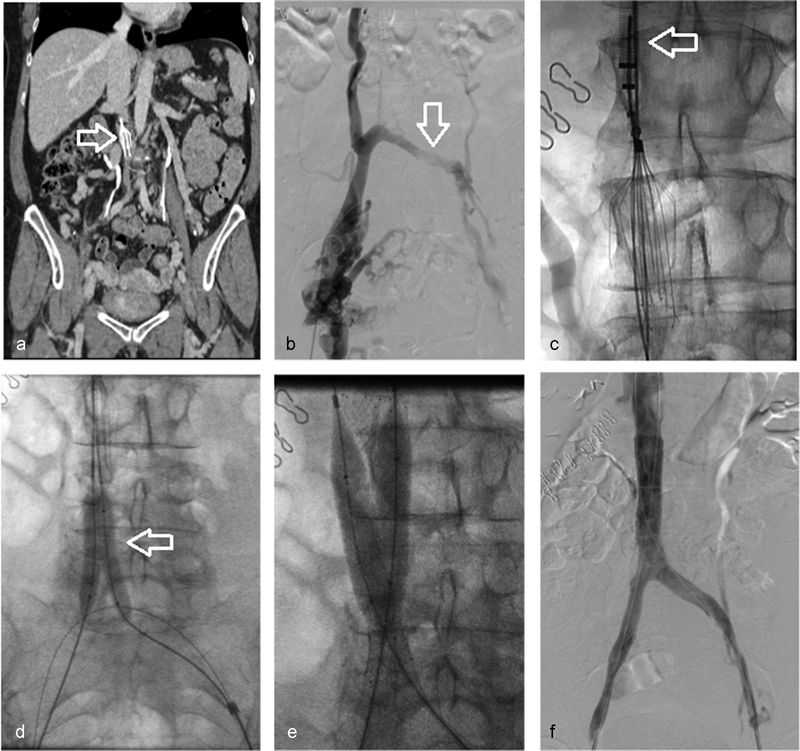
Successful balloon venoplasty and stenting in a 58-year-old woman with chronic thrombosis of the inferior vena cava and bilateral iliac veins after Denali filter (Bard Peripheral Vascular, Tempe, AZ) placement. (a) CT venogram demonstrating occlusion of the IVC and opacification of multiple venous collaterals (arrow—IVC filter). (b) Venography confirms occlusion of the left external iliac vein with mature collaterals draining across the pelvis and into the IVC via lumbar veni collaterals (arrow). (c) Via internal jugular access, an endovascular snare was utilized to capture the filter hook. The filter was then coaxially collapsed in the sheaths (arrow). (d) Sequential 8-mm balloon angioplasty (arrow) of the iliac veins and IVC was performed from each access. (e) 14 mm × 80 mm self-expanding stents were deployed into the IVC immediately cranial to the prior location of the filter. (f) Completion venography demonstrating flow through the stented segments (arrows) without evidence of residual flow-limiting lesion.
Similar articles
-
Presentation and Management of Inferior Vena Cava Thrombosis.Ann Vasc Surg. 2019 Apr;56:17-23. doi: 10.1016/j.avsg.2018.08.082. Epub 2018 Oct 27. Ann Vasc Surg. 2019. PMID: 30982504
-
Efficacy of Retrievable Inferior Vena Cava Filter Placement in the Prevention of Pulmonary Embolism during Catheter-Directed Thrombectomy for Proximal Lower-Extremity Deep Vein Thrombosis.Ann Vasc Surg. 2016 May;33:181-6. doi: 10.1016/j.avsg.2015.10.034. Epub 2016 Jan 22. Ann Vasc Surg. 2016. PMID: 26806235
-
Catheter directed interventions for inferior vena cava thrombosis.Cardiovasc Diagn Ther. 2016 Dec;6(6):612-622. doi: 10.21037/cdt.2016.11.09. Cardiovasc Diagn Ther. 2016. PMID: 28123981 Free PMC article. Review.
-
Pharmacomechanical thrombolysis in the management of acute inferior vena cava filter occlusion using the Trellis-8 device.J Endovasc Ther. 2015 Feb;22(1):99-104. doi: 10.1177/1526602814564369. J Endovasc Ther. 2015. PMID: 25775688
-
Inferior Vena Cava Thrombosis.JACC Cardiovasc Interv. 2016 Apr 11;9(7):629-43. doi: 10.1016/j.jcin.2015.12.268. Epub 2016 Mar 4. JACC Cardiovasc Interv. 2016. PMID: 26952909 Review.
Cited by
-
Obstructive Shock in Acute Vena Cava Filter Thrombosis: A Rare Presentation.Clin Med Res. 2024 Sep;22(3):156-159. doi: 10.3121/cmr.2024.1921. Clin Med Res. 2024. PMID: 39438149 Free PMC article.
-
IVC filter use in patients with a history of venous thromboembolism undergoing bariatric surgery: a MBSAQIP study.Surg Endosc. 2025 Feb;39(2):875-880. doi: 10.1007/s00464-024-11395-5. Epub 2024 Dec 2. Surg Endosc. 2025. PMID: 39623172
-
Protrieve Sheath embolic protection during venous thrombectomy: early experience in seventeen patients.CVIR Endovasc. 2024 Oct 9;7(1):74. doi: 10.1186/s42155-024-00484-0. CVIR Endovasc. 2024. PMID: 39382712 Free PMC article.
-
Recurrent Deep Vein Thrombosis (DVT) and Inferior Vena Cava (IVC) Filter: Should a Computed Tomography (CT) Venogram and Inferior Vena Cavagram Be the Standard of Care?Cureus. 2024 Apr 18;16(4):e58529. doi: 10.7759/cureus.58529. eCollection 2024 Apr. Cureus. 2024. PMID: 38957832 Free PMC article.
-
Thrombogenic and Inflammatory Reactions to Biomaterials in Medical Devices.Front Bioeng Biotechnol. 2020 Mar 12;8:123. doi: 10.3389/fbioe.2020.00123. eCollection 2020. Front Bioeng Biotechnol. 2020. PMID: 32226783 Free PMC article. Review.
References
-
- Geerts W H Bergqvist D Pineo G F et al.Prevention of venous thromboembolism: American College of Chest Physicians Evidence-Based Clinical Practice Guidelines (8th Edition) Chest 2008133(6, Suppl):381S–453S. - PubMed
-
- Kaufman J A, Kinney T B, Streiff M B. et al.Guidelines for the use of retrievable and convertible vena cava filters: report from the Society of Interventional Radiology multidisciplinary consensus conference. J Vasc Interv Radiol. 2006;17(3):449–459. - PubMed
-
- PREPIC Study Group . Eight-year follow-up of patients with permanent vena cava filters in the prevention of pulmonary embolism: the PREPIC (Prevention du Risque d'Embolie Pulmonaire par Interruption Cave) randomized study. Circulation. 2005;112(3):416–422. - PubMed
-
- Decousus H, Leizorovicz A, Parent F. et al.A clinical trial of vena caval filters in the prevention of pulmonary embolism in patients with proximal deep-vein thrombosis. Prévention du Risque d'Embolie Pulmonaire par Interruption Cave Study Group. N Engl J Med. 1998;338(7):409–415. - PubMed
-
- Rajasekhar A, Streiff M B. Vena cava filters for management of venous thromboembolism: a clinical review. Blood Rev. 2013;27(5):225–241. - PubMed
Publication types
LinkOut - more resources
Full Text Sources
Other Literature Sources
