

Incremental value of thoracic ultrasound in intensive care units: Indications, uses, and applications
- PMID: 27247712
- PMCID: PMC4882403
- DOI: 10.4329/wjr.v8.i5.460
Incremental value of thoracic ultrasound in intensive care units: Indications, uses, and applications
Abstract
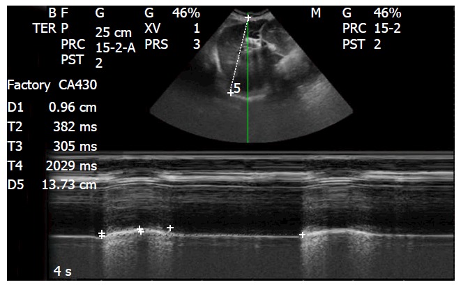
Emergency physicians are required to care for unstable patients with life-threatening conditions, and thus must make decisions that are both quick and precise about unclear clinical situations. There is increasing consensus in favor of using ultrasound as a real-time bedside clinical tool for clinicians in emergency settings alongside the irreplaceable use of historical and physical examinations. B-mode sonography is an old technology that was first proposed for medical applications more than 50 years ago. Its application in the diagnosis of thoracic diseases has always been considered limited, due to the presence of air in the lung and the presence of the bones of the thoracic cage, which prevent the progression of the ultrasound beam. However, the close relationship between air and water in the lungs causes a variety of artifacts on ultrasounds. At the bedside, thoracic ultrasound is based primarily on the analysis of these artifacts, with the aim of improving accuracy and safety in the diagnosis and therapy of the various varieties of pulmonary pathologic diseases which are predominantly "water-rich" or "air-rich". The indications, contraindications, advantages, disadvantages, and techniques of thoracic ultrasound and its related procedures are analyzed in the present review.
Keywords: Echocardiography; Heart failure; Intensive care unit; Pleural effusion; Pneumothorax; Thoracic ultrasound.
Figures

Similar articles
-
Lung sonography.J Ultrasound Med. 2013 Jan;32(1):165-71. doi: 10.7863/jum.2013.32.1.165. J Ultrasound Med. 2013. PMID: 23269722 Review.
-
Point-of-care lung ultrasound.Praxis (Bern 1994). 2014 Jun 4;103(12):711-6. doi: 10.1024/1661-8157/a001690. Praxis (Bern 1994). 2014. PMID: 24894615 Review.
-
Is thoracic ultrasound a viable alternative to conventional imaging in the critical care setting?Br J Anaesth. 2013 Aug;111(2):152-60. doi: 10.1093/bja/aet076. Epub 2013 Apr 12. Br J Anaesth. 2013. PMID: 23585400 Review.
-
Ultrasound examination of the lungs in the intensive care unit.Pediatr Crit Care Med. 2009 Nov;10(6):693-8. doi: 10.1097/PCC.0b013e3181b7f637. Pediatr Crit Care Med. 2009. PMID: 19675509 Review.
-
[Value of lung ultrasound in emergency and intensive care medicine].Dtsch Med Wochenschr. 2014 Nov;139(45):2301-7. doi: 10.1055/s-0034-1387309. Epub 2014 Oct 28. Dtsch Med Wochenschr. 2014. PMID: 25350245 Review. German.
Cited by
-
[Emergency lung ultrasound].Med Klin Intensivmed Notfmed. 2018 Nov;113(8):616-624. doi: 10.1007/s00063-018-0485-z. Epub 2018 Oct 10. Med Klin Intensivmed Notfmed. 2018. PMID: 30306189 Review. German.
-
The PIEPEAR Workflow: A Critical Care Ultrasound Based 7-Step Approach as a Standard Procedure to Manage Patients with Acute Cardiorespiratory Compromise, with Two Example Cases Presented.Biomed Res Int. 2018 Jun 11;2018:4687346. doi: 10.1155/2018/4687346. eCollection 2018. Biomed Res Int. 2018. PMID: 29992144 Free PMC article.
-
Thoracic ultrasound: An adjunctive and valuable imaging tool in emergency, resource-limited settings and for a sustainable monitoring of patients.World J Radiol. 2016 Sep 28;8(9):775-784. doi: 10.4329/wjr.v8.i9.775. World J Radiol. 2016. PMID: 27721940 Free PMC article. Review.
-
Poor lung ultrasound score in shock patients admitted to the ICU is associated with worse outcome.BMC Pulm Med. 2019 Jan 3;19(1):1. doi: 10.1186/s12890-018-0755-9. BMC Pulm Med. 2019. PMID: 30606165 Free PMC article.
-
Bedside Ultrasound for Hemodynamic Monitoring in Cardiac Intensive Care Unit.J Clin Med. 2022 Dec 19;11(24):7538. doi: 10.3390/jcm11247538. J Clin Med. 2022. PMID: 36556154 Free PMC article. Review.
References
-
- Elia F, Panero F, Molino P, Ferrari G, Aprà F. Ultrasound to reduce cognitive errors in the ED. Am J Emerg Med. 2012;30:2030–2033. - PubMed
-
- American College of Emergency Physicians. Emergency ultrasound guidelines. Ann Emerg Med. 2009;53:550–570. - PubMed
-
- Volpicelli G, Elbarbary M, Blaivas M, Lichtenstein DA, Mathis G, Kirkpatrick AW, Melniker L, Gargani L, Noble VE, Via G, et al. International evidence-based recommendations for point-of-care lung ultrasound. Intensive Care Med. 2012;38:577–591. - PubMed
-
- Bekemeyer WB, Crapo RO, Calhoon S, Cannon CY, Clayton PD. Efficacy of chest radiography in a respiratory intensive care unit. A prospective study. Chest. 1985;88:691–696. - PubMed
-
- Zanobetti M, Poggioni C, Pini R. Can chest ultrasonography replace standard chest radiography for evaluation of acute dyspnea in the ED? Chest. 2011;139:1140–1147. - PubMed
Publication types
LinkOut - more resources
Full Text Sources
Other Literature Sources
