. 2016 May 12:5:610.
doi: 10.1186/s40064-016-2281-7. eCollection 2016.

The protective effect of hydromorphone to ischemia in rat glial cells

Affiliations

The protective effect of hydromorphone to ischemia in rat glial cells

Young Sung Kim et al. Springerplus. .

Abstract

Background: Ischemic insults during operation can cause ischemic-reperfusion injuries in brain as well as memory impairments. Total intravenous anesthesia (TIVA) is the preferred anesthetic method in brain surgery, as it utilizes motor evoked potential monitoring. And the use of opioids is common in TIVA. However there are few studies about ischemic protective effect of opioids to glial cells.

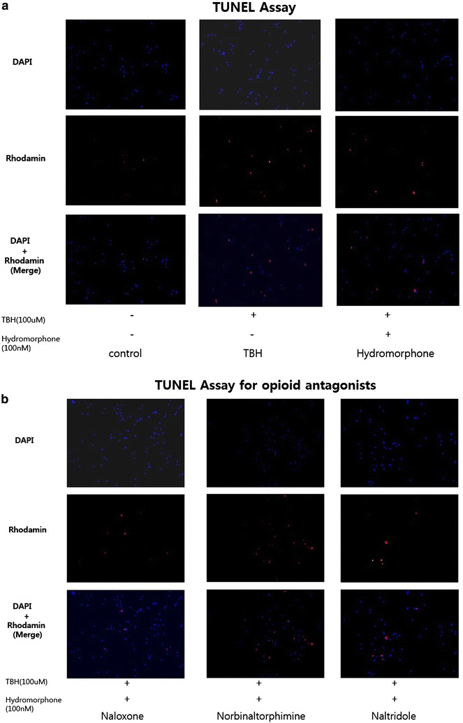
Methods: We used mixed cultures of rat glial cells, which were harvested from the brain of 1-day old rat. We divided the experimental groups according to their hydromorphone conditioning period: (a) pre-culture, (b) per-culture, or (c) pre- and per-culture. We measured the levels of the reactive oxygen species (ROS) induced by tert-butyl hydroperoxide (TBH) using flow cytometry. The ROS levels in the glial cells were also measured after the administration of 100 nM hydromorphone and selective opioid receptor antagonists.

Results: The ROS levels were reduced in the hydromorphone-treated group, as compared to the control group (only TBH treated). There were no differences between pre-conditioned and per-conditioned groups. However, the ROS levels were more reduced in pre- and per-conditioned group compared to pre-conditioned or per-conditioned only groups. Furthermore, selective antagonists for the delta, kappa, or mu opioid receptor partially negated the hydromorphone effect.

Conclusion: This study demonstrated that hydromorphone can have additional protective effects on oxidative stress when pre- and per-conditioning is combined. Furthermore we proved that μ, δ, κ opioid receptors participate in protective mechanism of hydromorphone to glial cells.

Keywords: Hydromorphone; Neuroglia; Reactive oxygen species.


Figures

Fig. 1
Fig. 1
Experimental design. TBH: tert-butyl hydroperoxide, DCF-DA: 2′,7′-dichloroflurorescin diacetate, OR antagonist : naltrindole (a selective delta opioid receptor antagonist) or nor-binaltorphimine (a selective kappa opioid receptor antagonist) or naloxone
Fig. 2
Fig. 2
This graph shows effective dosage of hydromorphone for reducing ROS. The amount of ROS was expressed as Mean Fluorescence index. *<0.05 versus TBH group. <0.05 versus 10 nM hydromorphone group
Fig. 3
Fig. 3
Detection of intracellular ROS (reactive oxygen species) product. This graph shows the protective effect of hydromorphone on TBH induced toxicity in primary rat glial cell cultures. The ROS level was measured with using FACS (DCF-DA) in the 100 nM hydromorphone (1) pre conditioned (2) per-conditioned (3) pre- and per- conditioned primary rat glial cells. The amount of ROS was expressed as Mean Fluorescence index. *<0.05 versus TBH group. <0.05 versus preconditioning group. <0.05 versus perconditioning group
Fig. 4
Fig. 4
Effects of opioid receptor (OR) antagonists on reactive oxygen species (ROS) products. This graph shows that selective OR antagonists reverse the attenuation of the ROS induced by hydromorphone (pre and per-condition). The ROS level was measured with using FACS (DCF-DA) in the cells treated only TBH (control group), and the cells treated with TBH, hydromorphone and/or one of selective delta, kappa, and mu opioid receptor antagonists, naltrindole, nor-binaltorphimine, and naloxone, respectively. The amount of ROS was expressed as Mean Fluorescence index.*<0.05 versus control group. +<0.05 versus hydromorphone group
Fig. 5
Fig. 5
Terminal deoxynucleotidyl transferase-mediated dUTP nick end labeling (TUNEL) assay of rat glial cells. a Fluorescence micrographs of cells were stained with rhodamine and DAPI. Red particles represent DNA fragments. This graphs show the Control, TBH, and Hydromorphone groups. b Fluorescence micrographs of cells were stained with rhodamine and DAPI. Red particles represent DNA fragments. This graphs show the opioid antagonist groups. <0.05 versus hydromorphone group
Fig. 6
Fig. 6
Terminal deoxynucleotidyl transferase-mediated dUTP nick end labeling (TUNEL) assay of rat glial cells. a This graph shows quantitation of apoptotic cells from TUNEL assay by number of red particles. *<0.05 versus TBH group. <0.05 versus hydromorphone group

References

    1. Askari N, Mahboudi F, Haeri-Rohani A, Kazemi B, Sarrami R, Edalat R, Ahmadiani A. Effects of single administration of morphine on G-protein mRNA level in the presence and absence of inflammation in the rat spinal cord. Scand J Immunol. 2008;67(1):47–52. doi: 10.1111/j.1365-3083.2007.02043.x. - DOI - PubMed
    1. Bilotta F, Gelb AW, Stazi E, Titi L, Paoloni FP, Rosa G. Pharmacological perioperative brain neuroprotection: a qualitative review of randomized clinical trials. Br J Anaesth. 2013;110(Suppl 1):i113–120. doi: 10.1093/bja/aet059. - DOI - PubMed
    1. Brown GC, Bal-Price A. Inflammatory neurodegeneration mediated by nitric oxide, glutamate, and mitochondria. Mol Neurobiol. 2003;27(3):325–355. doi: 10.1385/MN:27:3:325. - DOI - PubMed
    1. Calvo CF, Cesselin F, Gelman M, Glowinski J. Identification of an opioid peptide secreted by rat embryonic mixed brain cells as a promoter of macrophage migration. Eur J Neurosci. 2000;12:2676–2684. doi: 10.1046/j.1460-9568.2000.00145.x. - DOI - PubMed
    1. Das M, Das DK. Molecular mechanism of preconditioning. IUBMB Life. 2008;60(4):199–203. doi: 10.1002/iub.31. - DOI - PubMed

LinkOut - more resources