Review
. 2016:2016:9375753.
doi: 10.1155/2016/9375753. Epub 2016 May 9.

Practical Application of Columbia Classification for Focal Segmental Glomerulosclerosis

Affiliations
Review

Practical Application of Columbia Classification for Focal Segmental Glomerulosclerosis

Man-Hoon Han et al. Biomed Res Int. 2016.

Abstract

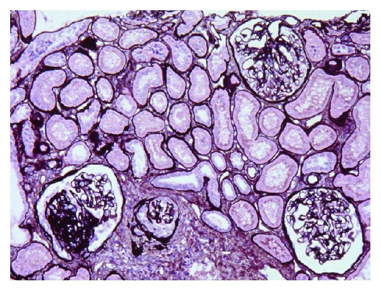
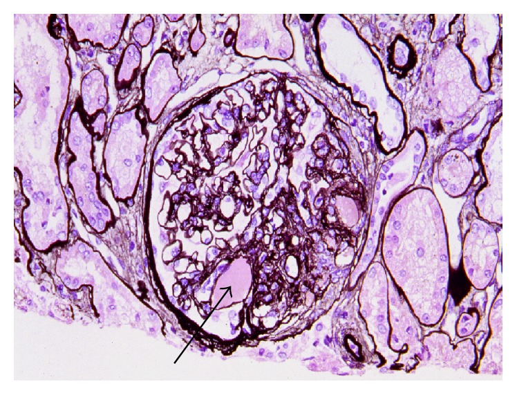
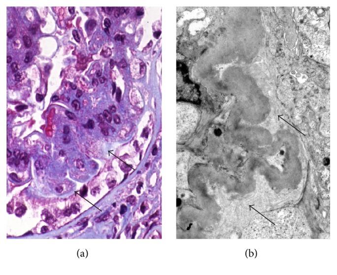
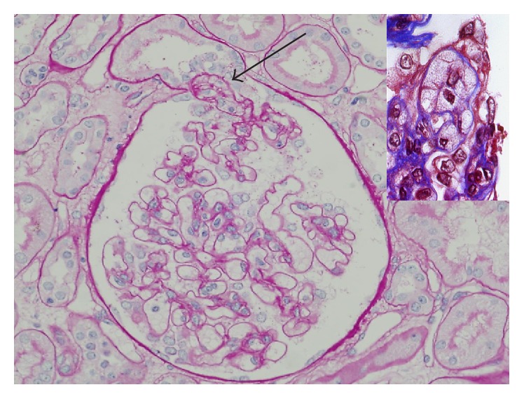
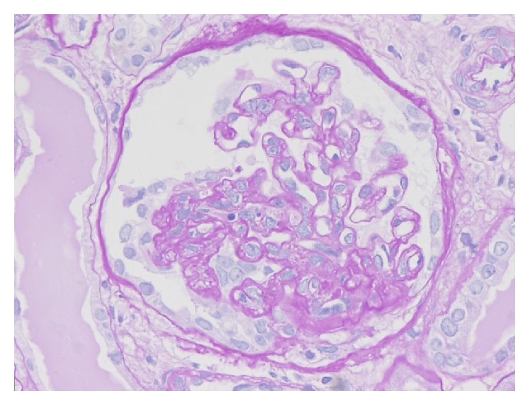
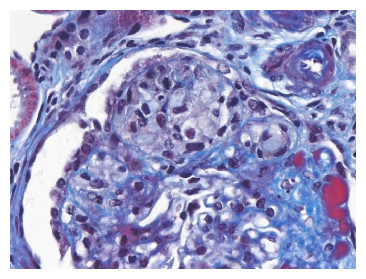
Focal segmental glomerulosclerosis (FSGS) is a heterogeneous clinicopathological entity. Two frameworks for the classification of FSGS have been described: etiologic and morphologic. The etiologic classification is distinguished among genetic, adaptive, virus-associated, drug-induced, and idiopathic types. Morphologic classification is commonly referred to as the Columbia classification published in 2004, which distinguishes five variants: collapsing, tip, cellular, perihilar, and not otherwise specified (NOS). This classification is based on light microscopic patterns with rigorously defined specific criteria, which can be applied to primary and secondary forms of FSGS, and has been widely used over the past 10 years both as a diagnostic and as a prognostic clinical tool. This paper defines common histopathological features of FSGS, distinguished characters among five variants, and points out the confusion about terminology of variants, because most were proposed in the past with different definitions. Despite good interobserver reproducibility of this classification system, difficulty in its application may arise in the interpretation of lesions with mixed features of more than one variant in the same tissue specimen and with late lesions, because other variants may evolve into the NOS variant over time.


Figures

Figure 1
Figure 1
Segmental sclerotic glomerulus is at left lower and right two glomeruli look normal. Interstitial fibrosis and tubular atrophy are observed focally. Artery shows intimal fibrosis. Jones methenamine silver stain (PAM), ×200. (Permitted by the Journal of Korean Society of Pediatric Nephrology [3].)
Figure 2
Figure 2
Perihilar variant of FSGS. Sclerosis is observed at the glomerular vascular pole. Hyaline (arrow) is the amorphous material in the middle of sclerosis, PAM, ×200. (Permitted by the Journal of Korean Society of Pediatric Nephrology [3].)
Figure 3
Figure 3
Halo formation. In trichrome stain (a), the pale zone (arrow) is between sclerosis and overlying podocytes. Trichrome stain, ×400. Some areas on the electron micrograph (b) are filled with newly formed thin collagen bundles (arrows). ×5000. (Permitted by the Journal of Korean Society of Pediatric Nephrology [3].)
Figure 4
Figure 4
Tip variant of FSGS. Foam cell accumulated segment prolapsed into the tubular pole, the origin of the proximal tubule. The remainder of the glomerular tuft appears normal. PAS stain, ×200. (Courtesy of Professor Mi Sun Choi, DongSan Hospital of Keymyung Medical College, Daegu, Korea.) Inset shows endocapillary foam cells in trichrome stain. ×400.
Figure 5
Figure 5
Collapsing variant of FSGS. Segmental collapse of glomerular capillaries is accompanied by proliferation of overlying podocytes. PAS stain, ×200.
Figure 6
Figure 6
Cellular variant of FSGS. Segment is expanded by endocapillary foam cells. Overlying epithelial cells are also prominent, but capillary collapse is not observed. Trichrome stain, ×200.

References

    1. Jennette J. C., Olson J. L., Schwartz M. M., Silva F. G. Heptinstall's Pathology of the Kidney. 6th. Philadelphia, Pa, USA: Lippincott Williams & Wilkins; 2007.
    1. Stokes M. B., D'Agati V. D. Morphologic variants of focal segmental glomerulosclerosis and their significance. Advances in Chronic Kidney Disease. 2014;21(5):400–407. doi: 10.1053/j.ackd.2014.02.010. - DOI - PubMed
    1. Kim Y. J. Pathology and classification of focal segmental glomerulosclerosis. Journal of the Korean Society of Pediatric Nephrology. 2012;16(1):21–31. doi: 10.3339/jkspn.2012.16.1.21. - DOI
    1. Newbold K. M., Sandison A., Howie A. J. Comparison of size of juxtamedullary and outer cortical glomeruli in normal adult kidney. Virchows Archiv A: Pathological Anatomy and Histopathology. 1992;420(2):127–129. doi: 10.1007/bf02358803. - DOI - PubMed
    1. Skov K., Nyengaard J. R., Patwardan A., Mulvany M. J. Large juxtamedullary glomeruli and afferent arterioles in healthy primates. Kidney International. 1999;55(4):1462–1469. doi: 10.1046/j.1523-1755.1999.00398.x. - DOI - PubMed

MeSH terms

LinkOut - more resources