. 2016 Feb 28;4(3):344-58.
doi: 10.1002/mgg3.208. eCollection 2016 May.

Analysis of a large choroideremia dataset does not suggest a preference for inclusion of certain genotypes in future trials of gene therapy

Affiliations

Analysis of a large choroideremia dataset does not suggest a preference for inclusion of certain genotypes in future trials of gene therapy

Paul R Freund et al. Mol Genet Genomic Med. .

Abstract

Background: Choroideremia (CHM) is an X-linked degeneration of the retinal pigment epithelium, photoreceptors, and choroid, which causes nyctalopia and progressive constriction of visual fields leading to blindness. The CHM gene encodes Rab escort protein 1 (REP-1). In this work, we reviewed the phenotypes and genotypes of affected males with the purpose of understanding the functional effects of CHM mutations and their relationship with the phenotypes.

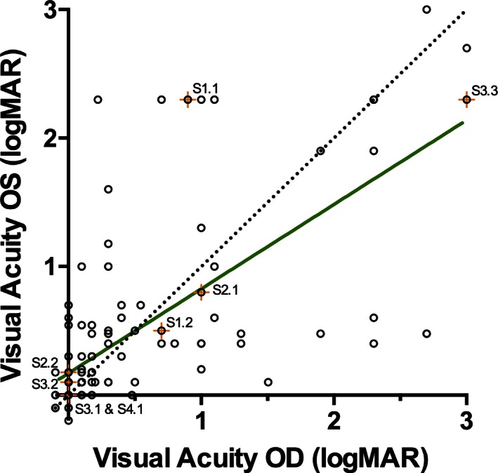
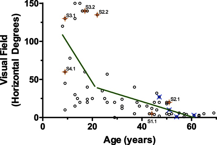
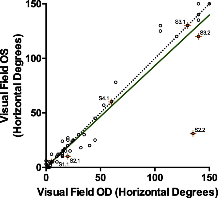
Methods: A retrospective review of 128 affected males was performed analyzing the onset of symptoms, visual acuity, and visual fields with respect to their mutations in the CHM gene.

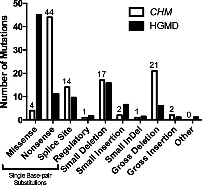
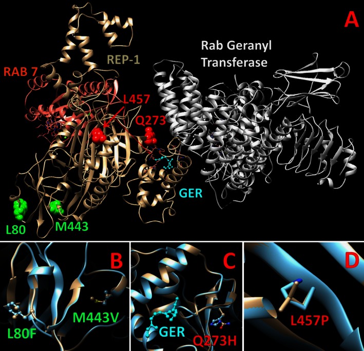
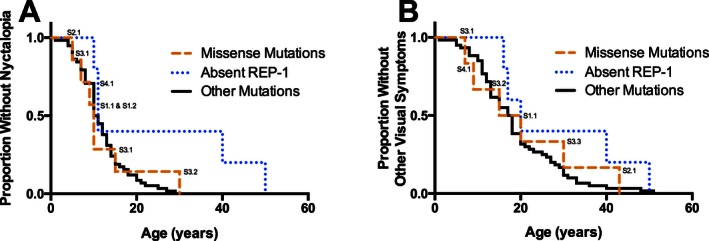
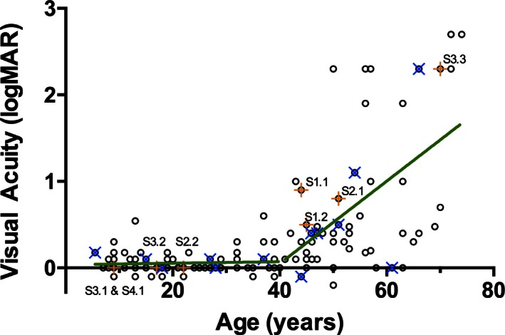
Results: In rank order, reflecting data from this report, the most common mutations found in the CHM gene were nonsense mutations (41%), exon deletions (37%), and splice sites (14%) associated with a loss of functional protein. In the pool of 106 CHM mutations, we discovered four novel missense mutations (c.238C>T; p.L80F, c.819G>T; p.Q273H, c.1327A>G; p.M443V, and c.1370C>T; p.L457P) predicted to be severe changes affecting protein stability and folding with the effect similar to that of other types of mutations. No significant genotype-phenotype correlation was found with respect to the onset of nyctalopia, the onset of other visual symptoms, visual acuity, or width of visual fields.

Conclusion: There is no evidence to support exclusion of CHM patients from clinical trials based on their genotypes or any potential genotype-phenotype correlations.

Keywords: Choroideremia; Rab escort protein‐1; natural history; retinal dystrophy; visual acuity; visual fields.


Figures

Figure 1
Figure 1
Proportion of CHM (MIM# 303100) gene mutations observed in 106 affected families compared to the mutations reported in the Human Genome Mutation Database (HGMD).
Figure 2
Figure 2
The structure of Rab escort protein 1 (REP‐1; beige) in relation to Rab geranyl transferase (white) and Ras‐associated protein 7 (RAB 7; pink) and its substrate geranylgeranyl (GER; blue). The REP‐1 double mutant, L80F and M443V, is highlighted in panel B (green); the Q273H (red) and L457P (red) mutations are in panels C and D, respectively.
Figure 3
Figure 3
Kaplan–Meier survival curves demonstrating the self‐reported onset (in years) of nyctalopia (A) or other visual symptoms (B) in males affected by choroideremia. Subjects are grouped by the causative mutation: subjects with missense mutations (labeled; median age of onset of nyctalopia = 10 years, n = 7; median age of onset of other symptoms = 17.5 years, n = 6), subjects who do not express any Rab escort protein 1 due to whole gene deletions or deletions of the ATG start codon (median age of onset of nyctalopia = 11 years, n = 5; median age of onset of other symptoms = 20 years, n = 5), and subjects with other disease causing mutations (median age of onset of nyctalopia = 10.5 years, n = 58; onset of other symptoms = 17.5 years, n = 60). There is no significant difference between the survival curves of the three groups (nyctalopia: P = 0.08; other visual symptoms: P = 0.35).
Figure 4
Figure 4
The best‐corrected visual acuity (logMAR equivalent) of affected males' better eye as a function of age (n = 128). The sample population is divided at the critical age (40 years old) into the ≤40 years old group or the >40 years old group. Individuals with missense mutations are indicated by an orange + and labeled; individuals who do not express any Rab escort protein 1 due to whole gene deletions or deletions of the ATG start codon are indicated by a blue ×. The rate of change of visual acuity predicted by a linear regression model (green line) in the ≤40 years old group is not significantly different from 0 (P = 0.71); the rate of change of visual acuity in the >40 years old group is 0.0483 logMAR units/year (P = 0.001).
Figure 5
Figure 5
The intereye correlation of the best‐corrected visual acuities (logMAR) of affected males. A perfect correlation is also plotted for comparison (dotted line). The visual acuities of individuals' eyes are highly correlated (Spearman r = 0.76). Subjects with missense mutations are indicated by an orange + and labeled. OS, left eye; OD, right eye
Figure 6
Figure 6
The visual field (the continuous visual field across the horizontal meridian, in degrees) of affected males' better eye as a function of age (n = 64). The sample population was divided at the critical age (20 years old), separating individuals into the ≤20 years old group and the >20 years old group. Individuals with missense mutations are indicated by an orange + and labeled; individuals who do not express any Rab escort protein 1 due to whole gene deletions or deletions of the ATG start codon are indicated by a blue ×. In a linear regression model (green line), age was not a significant predictor of visual field in the ≤20 years old group (P = 0.785); the rate of change of visual fields in the >20 years old group is a loss of 0.868 horizontal degrees per year (P = 0.005).
Figure 7
Figure 7
The intereye correlation of the visual fields (horizontal degrees across the meridian) of affected males. A perfect correlation is plotted for comparison (dotted line). The visual fields of individuals' eyes are very highly correlated (Spearman r = 0.95). Subjects with missense mutations are indicated by an orange + and labeled. OS, left eye; OD, right eye.

Similar articles

Cited by

References

    1. Abstracts of the 50th ISCEV (International Society for Clinical Electrophysiology of Vision) International Symposium, June 3–7, 2012, Valencia, Spain . 2012. Doc. Ophthalmol. 124(Suppl 1):1–68. - PubMed
    1. Beaufrere, L. , Claustres M., and Tuffery S.. 1999. No missense mutation in choroideremia patients analyzed to date. Ophthalmic Genet. 20:89–93. - PubMed
    1. Bentley, D. R. , Balasubramanian S., Swerdlow H. P., Smith G. P., Milton J., Brown C. G., et al. , et al. 2008. Accurate whole human genome sequencing using reversible terminator chemistry. Nature 456:53–59. - PMC - PubMed
    1. Berson, E. L. , Sandberg M. A., Rosner B., Birch D. G., and Hanson A. H.. 1985. Natural course of retinitis pigmentosa over a three‐year interval. Am. J. Ophthalmol. 99:240–251. - PubMed
    1. Bittner, A. K. , Iftikhar M. H., and Dagnelie G.. 2011. Test‐retest, within‐visit variability of goldmann visual fields in retinitis pigmentosa. Invest. Ophthalmol. Vis. Sci. 52:8042–8046. - PMC - PubMed

LinkOut - more resources