

Prions, amyloids, and RNA: Pieces of a puzzle
- PMID: 27248002
- PMCID: PMC4981203
- DOI: 10.1080/19336896.2016.1181253
Prions, amyloids, and RNA: Pieces of a puzzle
Abstract
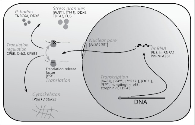
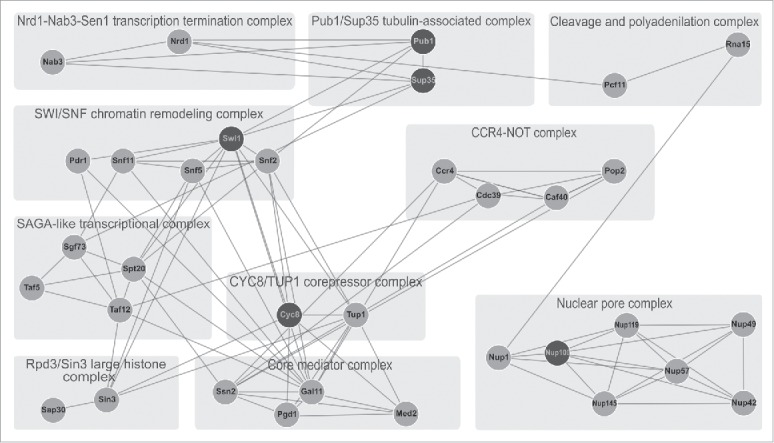
Amyloids are protein aggregates consisting of fibrils rich in β-sheets. Growth of amyloid fibrils occurs by the addition of protein molecules to the tip of an aggregate with a concurrent change of a conformation. Thus, amyloids are self-propagating protein conformations. In certain cases these conformations are transmissible / infectious; they are known as prions. Initially, amyloids were discovered as pathological extracellular deposits occurring in different tissues and organs. To date, amyloids and prions have been associated with over 30 incurable diseases in humans and animals. However, a number of recent studies demonstrate that amyloids are also functionally involved in a variety of biological processes, from biofilm formation by bacteria, to long-term memory in animals. Interestingly, amyloid-forming proteins are highly overrepresented among cellular factors engaged in all stages of mRNA life cycle: from transcription and translation, to storage and degradation. Here we review rapidly accumulating data on functional and pathogenic amyloids associated with mRNA processing, and discuss possible significance of prion and amyloid networks in the modulation of key cellular functions.
Keywords: Amyloid; CPEB; Prion; Pub1; S. cerevisiae; Sup35; Tia1; yeast.
Figures
Similar articles
-
[Yeast prions, mammalian amyloidoses, and the problem of proteomic networks].Genetika. 2006 Nov;42(11):1558-70. Genetika. 2006. PMID: 17163073 Review. Russian.
-
Amyloid properties of the yeast cell wall protein Toh1 and its interaction with prion proteins Rnq1 and Sup35.Prion. 2019 Jan;13(1):21-32. doi: 10.1080/19336896.2018.1558763. Epub 2018 Dec 27. Prion. 2019. PMID: 30558459 Free PMC article.
-
Application of yeast to studying amyloid and prion diseases.Adv Genet. 2020;105:293-380. doi: 10.1016/bs.adgen.2020.01.002. Epub 2020 May 4. Adv Genet. 2020. PMID: 32560789 Free PMC article. Review.
-
Amyloids, prions and the inherent infectious nature of misfolded protein aggregates.Trends Biochem Sci. 2006 Mar;31(3):150-5. doi: 10.1016/j.tibs.2006.01.002. Epub 2006 Feb 13. Trends Biochem Sci. 2006. PMID: 16473510 Review.
-
Prions, prionoid complexes and amyloids: the bad, the good and something in between.Swiss Med Wkly. 2017 Apr 18;147:w14424. doi: 10.4414/smw.2017.14424. eCollection 2017. Swiss Med Wkly. 2017. PMID: 28421568 Review.
Cited by
-
RNA Sequencing Reveals Specific TranscriptomicSignatures Distinguishing Effects of the [SWI⁺] Prion and SWI1 Deletion in Yeast Saccharomyces cerevisiae.Genes (Basel). 2019 Mar 12;10(3):212. doi: 10.3390/genes10030212. Genes (Basel). 2019. PMID: 30871095 Free PMC article.
-
Functional Mammalian Amyloids and Amyloid-Like Proteins.Life (Basel). 2020 Aug 21;10(9):156. doi: 10.3390/life10090156. Life (Basel). 2020. PMID: 32825636 Free PMC article. Review.
-
RNA-binding protein FXR1 is presented in rat brain in amyloid form.Sci Rep. 2019 Dec 12;9(1):18983. doi: 10.1038/s41598-019-55528-6. Sci Rep. 2019. PMID: 31831836 Free PMC article.
-
Predicting Amyloidogenic Proteins in the Proteomes of Plants.Int J Mol Sci. 2017 Oct 16;18(10):2155. doi: 10.3390/ijms18102155. Int J Mol Sci. 2017. PMID: 29035294 Free PMC article.
-
Human cyclophilin 40 unravels neurotoxic amyloids.PLoS Biol. 2017 Jun 27;15(6):e2001336. doi: 10.1371/journal.pbio.2001336. eCollection 2017 Jun. PLoS Biol. 2017. PMID: 28654636 Free PMC article.
References
-
- Kyle RA. Amyloidosis: a convoluted story. Br J Haematol 2001; 114:529-38; PMID:11552976; http://dx.doi.org/10.1046/j.1365-2141.2001.02999.x - DOI - PubMed
-
- Buxbaum JN, Linke RP. A molecular history of the amyloidoses. J Mol Biol 2012; 421:142-59; PMID:22321796; http://dx.doi.org/10.1016/j.jmb.2012.01.024 - DOI - PubMed
-
- Virchow R. Ueber eine im Gehirn und Ruckenmark des Menschen aufgefunde Substanz mit der chemishen Reaction der Cellulose. Virchows Arch Path Anat Physiol 1854; 6:135-8; http://dx.doi.org/10.1007/BF01930815 - DOI
-
- Friedreich N, Kekule FA. Zur Amyloidfrage. Virchows Arch Path Anat Physiol 1859; 16:50-65; http://dx.doi.org/10.1007/BF01945246 - DOI
-
- Snow AD, Willmer J, Kisilevsky R. Sulfated glycosaminoglycans: a common constituent of all amyloids? Lab Invest 1987; 56:120-3; PMID:2432352 - PubMed
Publication types
MeSH terms
Substances
LinkOut - more resources
Full Text Sources
Other Literature Sources
Molecular Biology Databases
Miscellaneous