

Clusterin levels are increased in Alzheimer's disease and influence the regional distribution of Aβ
- PMID: 27248362
- PMCID: PMC8029153
- DOI: 10.1111/bpa.12392
Clusterin levels are increased in Alzheimer's disease and influence the regional distribution of Aβ
Abstract
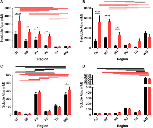
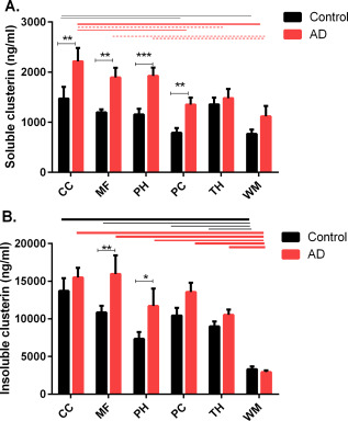
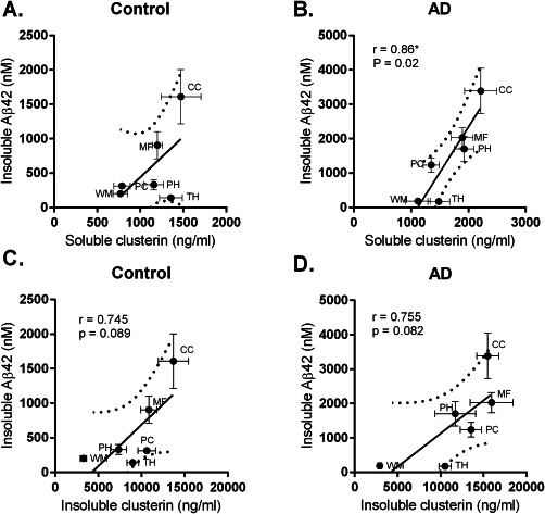
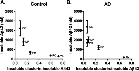
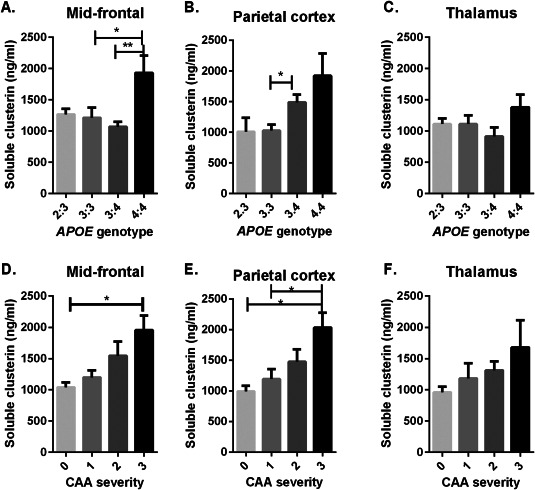
Clusterin, also known as apoJ, is a lipoprotein abundantly expressed within the CNS. It regulates Aβ fibril formation and toxicity and facilitates amyloid-β (Aβ) transport across the blood-brain barrier. Genome-wide association studies have shown variations in the clusterin gene (CLU) to influence the risk of developing sporadic Alzheimer's disease (AD). To explore whether clusterin modulates the regional deposition of Aβ, we measured levels of soluble (NP40-extracted) and insoluble (guanidine-HCl-extracted) clusterin, Aβ40 and Aβ42 by sandwich ELISA in brain regions with a predilection for amyloid pathology-mid-frontal cortex (MF), cingulate cortex (CC), parahippocampal cortex (PH), and regions with little or no pathology-thalamus (TH) and white matter (WM). Clusterin level was highest in regions with plaque pathology (MF, CC, PH and PC), approximately mirroring the regional distribution of Aβ. It was significantly higher in AD than controls, and correlated positively with Aβ42 and insoluble Aβ40. Soluble clusterin level rose significantly with severity of cerebral amyloid angiopathy, and in MF and PC regions was highest in APOE ɛ4 homozygotes. In the TH and WM (areas with little amyloid pathology) clusterin was unaltered in AD and did not correlate with Aβ level. There was a significant positive correlation between the concentration of clusterin and the regional levels of insoluble Aβ42; however, the molar ratio of clusterin : Aβ42 declined with insoluble Aβ42 level in a region-dependent manner, being lowest in regions with predilection for Aβ plaque pathology. Under physiological conditions, clusterin reduces aggregation and promotes clearance of Aβ. Our findings indicate that in AD, clusterin increases, particularly in regions with most abundant Aβ, but because the increase does not match the rising level of Aβ42, the molar ratio of clusterin : Aβ42 in those regions falls, probably contributing to Aβ deposition within the tissue.
Keywords: Alzheimer's disease; amyloid-β; amyloid-β clearance; apoJ; cerebral amyloid angiopathy; clusterin; plaque.
© 2016 International Society of Neuropathology.
Figures
References
-
- Hardy J, Allsop D (1991) Amyloid deposition as the central event in the aetiology of Alzheimer's disease. Trends Pharmacol Sci 12:383–388. - PubMed
-
- Gravina SA, Ho L, Eckman CB, Long KE, Otvos L, Jr ., Younkin LH, et al (1995) Amyloid β protein (Aβ) in Alzheimer's disease brain. Biochemical and immunocytochemical analysis with antibodies specific for forms ending at Aβ40 or Aβ42(43). J Biol Chem 270:7013–7016. - PubMed
-
- Iwatsubo T, Odaka A, Suzuki N, Mizusawa H, Nukina N, Ihara Y (1994) Visualization of Aβ42(43) and Aβ40 in senile plaques with end‐specific Aβ monoclonals: Evidence that an initially deposited species is Aβ42(43). Neuron 13:45–53. - PubMed
-
- Price DL, Sisodia SS (1998) Mutant genes in familial Alzheimer's disease and transgenic models. Annu Rev Neurosci 21:479–505. - PubMed
Publication types
MeSH terms
Substances
Grants and funding
LinkOut - more resources
Full Text Sources
Other Literature Sources
Medical
Research Materials
Miscellaneous
