. 2016 Jun 1;11(6):e0156032.
doi: 10.1371/journal.pone.0156032. eCollection 2016.

A GCN2-Like eIF2α Kinase (LdeK1) of Leishmania donovani and Its Possible Role in Stress Response

Affiliations

A GCN2-Like eIF2α Kinase (LdeK1) of Leishmania donovani and Its Possible Role in Stress Response

Shilpa J Rao et al. PLoS One. .

Abstract

Translation regulation in Leishmania parasites assumes significance particularly because they encounter myriad of stresses during their life cycle. The eukaryotic initiation factor 2α (eIF2α) kinases, the well-known regulators of translation initiation in higher eukaryotes have now been found to control various processes in these protozoan parasites as well. Here, we report on cloning and characterization of a GCN2-like eIF2α kinase from L. donovani and also on its modulation during nutrient starvation. We cloned a GCN2-like kinase from L. donovani, which we named as LdeK1 and validated it to be a functional eIF2α kinase by in vitro kinase assay. LdeK1 was found to be localized in the cytoplasm of the promastigotes with a five-fold higher expression in this stage of the parasite as compared to the axenic amastigotes. Phosphorylation of eIF2α and a G1-arrest was observed in response to nutrient starvation in the wild-type parasites. In contrast, phosphorylation was significantly impaired in a dominant-negative mutant of LdeK1 during this stress with a subsequent failure to bring about a G1-arrest during cell cycle. Thus, LdeK1 is a functional GCN2-like kinase of L. donovani which responds to nutrient starvation by phosphorylating its substrate, eIF2α and a G1-arrest in the cell cycle. Nutrient starvation is encountered by the parasites inside the vector which triggers metacyclogenesis. We therefore propose that global translational regulation by activation of LdeK1 followed by eIF2α phosphorylation and G1-arrest during nutrient starvation in the gut of sandfly vector could be one of the mechanisms to retool the cellular machinery required for metacyclogenesis of Leishmania promastigotes.


Conflict of interest statement

Competing Interests: The authors have declared that no competing interests exist.

Figures

Fig 1
Fig 1. An eIF2α kinase (LdeK1) homologous to GCN2 is present in L. donovani.
(A) CLUSTAL W analysis of the catalytic domain of LdeK1 from L. donovani (KP325123) with GCN2 of Homo sapiens, HsGCN2 (NP_001013735); Mus musculus, MmGCN2 (NP_001171277); Saccharomyces cerevisiae, ScGCN2 (AAA34636); Drosophila melanogaster, DmGCN2 (AGB96521); Leishmania major, LmeK1 (XP_001681474); Trypanosoma brucei, TbK1, (XP_828792); Plasmodium falciparum, PfeIK1 (XP_001348597), Toxoplasma gondii, TgIF2K-C (AHM92904); TgIF2K-D (AED01979). The catalytic domain comprises of I-IX subdomains which are demarcated by solid black lines on top. Number between subdomain IV and V represents the variable length of the kinase insert present in different kinases. The kinase insert sequence has been omitted to facilitate the alignment. Identical residues are indicated in dark grey and similar sequences are highlighted in light grey. Dots represent residues conserved in kinases and shaded triangles indicate residues that are specific to eIF2α kinases. The arrow in subdomain VIII indicates the residue that is autophosphorylated during activation of PKR. (B) Domain organization of LdeK1 along with TgIF2K-D and MmGCN2 for comparison. The numbers demarcate the residue numbers for each domain of the proteins. (C) Phylogenetic analysis of LdeK1.
Fig 2
Fig 2. LdeK1 is a functional eIF2α kinase of L. donovani that phosphorylates LdeIF2α at T-166 residue in vitro.
The bacterially overexpressed catalytic domain of LdeK1 (LdeK1KD) was incubated with (A) wild-type LdeIF2α and LdeIF2α T166A or heIF2α (B) along with cold ATP at 30°C and subjected to SDS-PAGE. As a positive control, the bacterially overexpressed catalytic domain of human heme-regulated inhibitor (hHRIKD) was incubated with heIF2α along with cold ATP at 30°C and subjected to SDS-PAGE. The phosphorylation of eIF2α was analyzed by western blotting using anti-phospho-eIF2α antibody. (A) Leishmanial total eIF2α (LdeIF2α) and (B) human total eIF2α (heIF2α) were used as loading controls using Leishmania-specific and human-specific eIF2α antibodies, respectively. LdeK1 could phosphorylate LdeIF2α and not LdeF2αT166A indicating LdeK1 to be a functional eIF2α kinase capable of phosphorylating LdeIF2α at threonine-166 residue and heIF2α at serine-51 residue.
Fig 3
Fig 3. Expression of LdeK1 in L. donovani.
(A) LdeK1 is mainly expressed in the promastigotes of L. donovani at the mRNA level. Transcript levels of LdeK1 was analysed by quantitative real-time PCR in both L. donovani promastigotes and axenic amstigotes. The transcript level was approximately 5 fold more in promastigotes as compared to the axenic amastigotes.*p < 0.005 (B) Immunoblot to analyze the specificity of the antibody raised against LdeK1. The N-terminal domain of LdeK1 was bacterially overexpressed and partially purified followed by antibody production. The antibody could detect the source protein purified from E. coli and a protein at approximately 130 kDa in total leishmania lysates using western blotting. No protein was detected in Leishmania lysates with pre-immune sera and in MCF-7 lysates with immune sera, used to test the species specificity of the antibody. (C) Western blot analysis using anti-LdeK1 antibody revealed the expression of LdeK1 in the promastigote stage of the parasite. α-tubulin was used as a loading control.
Fig 4
Fig 4. LdeK1 is localized in the cytoplasm of L. donovani promastigotes.
(A.) Indirect immunofluorescence was performed using anti-LdeK1 antibody (immune-sera) and cy3-conjugated secondary antibody (red) to detect the localization of the LdeK1 in promastigotes of L. donovani. Nuclear DNA was stained with DAPI (blue). LdeK1 was found to localize in the cytoplasm of promastigotes. (B.) preimmune serum did not give any signal. (C.) negative control, stained with cy3-conjugated secondary antibody alone. (D) Cellular fractionation of promastigotes using digitonin resulted in a soluble cytosolic fraction (F1) and insoluble fraction including the organelle fraction (F2). The LdeK1 was detected in the F1 fraction by anti-LdeK1 antibody. α-tubulin was used as control for the cytoskeleton bound insoluble fraction.
Fig 5
Fig 5. Generation of dominant-negative mutant of L. donovani.
(A) Schematic representation of pIR1SAT vector used for generation of dominant negative mutant. The BglII was used for cloning of N- terminal domain of LdeK1. (B). Integration of the DNA fragment representing the N-terminal domain of LdeK1 in the genome of L. donovani. PCR using genomic DNA from untransfected and transfected (empty and test) L. donovani detected a 1000 bp band indicating the integration of the required fragment in the genome of L. donovani. (C) Overexpression of N-terminal segment of LdeK1 in L. donovani at mRNA-level. The overexpression of N-terminal segment of LdeK1 at the transcript level was analysed by RT-PCR which shows the overexpression in transfected Leishmania but not in Leishmania transfected with empty vector or untransfected Leishmania. (D) Overexpression of N-terminal domain of LdeK1 in L. donovani at protein level. The overexpression of N-terminal domain of LdeK1 at the protein level was analyzed by western blotting using anti-LdeK1 antibody which shows the overexpressed protein in transfected Leishmania [dominant negative mutant (DNM); lane 3] but not in Leishmania transfected with empty vector (lane2) or untransfected Leishmania (control; lane 1).
Fig 6
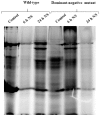
Fig 6. LdeK1 is involved during nutrient starvation to L. donovani.
(A) Both wild type and the dominant-negative mutant (DNM) parasites were subjected to nutrient starvation for 1 h, 2 h, 4 h, 6 h and 24 h in EBSS. Parasites starved for 4 h were also recovered in complete media for 24 h. Total protein lysates were subjected to SDS-PAGE and electrotransferred. The phosphorylation was analyzed by western blotting using phospho-eIF2α antibody. Phosphorylation of eIF2α occurs in response to nutrient starvation in wild-type L. donovani parasites. However it is not seen in case of dominant-negative mutants (DNM) indicating the role of LdeK1 in nutrient starvation condition. Total eIF2α was used as a loading control. (B) Densitometric analysis of the western blot profile (peIF2α/total eIF2α).
Fig 7
Fig 7. Cell cycle arrest in G1 phase during nutrient starvation is dependent on LdeK1 in L. donovani.
L. donovani promastigotes were subjected to nutrient starvation for 1 h, 2 h, 4 h, 6 h and 24 h followed by fixation in 70% methanol and PI staining. The stained parasites were analyzed for cell cycle distribution by FACS using 10000 events. Nutrient deprivation induced a G1-arrest in wild-type parasites. However, parasites lacking a functional LdeK1 did not show an arrest in this phase of cell-cycle, further suggesting its role in nutrient starvation condition in L. donovani.
Fig 8
Fig 8. De novo protein synthesis is affected in wild type parasites as compared to the dominant-negative mutant (DNM) during nutrient starvation.
Log-phase promastigotes both wild type and DNM were subjected to nutrient starvation for 6 h and 24 h followed by metabolic labelling of newly synthesized proteins using methionine analog L-azidohomoalanine (AHA). The protein synthesis is down regulated in the wild-type parasites at 6 h, however it is not affected in the DNM, indicating the role of LdeK1 in general protein synthesis.

References

    1. Sacks DL, Perkins P V. Identification of an infective stage of Leishmania promastigotes. Science. 1984;223: 1417–1419. 10.1126/science.6701528 - DOI - PubMed
    1. Da Silva R, Sacks DL. Metacyclogenesis is a major determinant of Leishmania promastigote virulence and attenuation. Infect Immun. 1987;55: 2802–2806. - PMC - PubMed
    1. Bates PA, Tetley L. Leishmania mexicana: induction of metacyclogenesis by cultivation of promastigotes at acidic pH. Exp Parasitol. 1993;76: 412–423. 10.1006/expr.1993.1050 - DOI - PubMed
    1. Serafim TD, Figueiredo AB, Costa PAC, Marques-da-Silva EA, Gonçalves R, de Moura SAL, et al. Leishmania Metacyclogenesis Is Promoted in the Absence of Purines. PLoS Negl Trop Dis. 2012;6 10.1371/journal.pntd.0001833 - DOI - PMC - PubMed
    1. Zilberstein D, Shapira M. The role of pH and temperature in the development of Leishmania parasites. Annu Rev Microbiol. 1994;48: 449–470. 10.1146/annurev.mi.48.100194.002313 - DOI - PubMed

LinkOut - more resources