Clinical Trial
. 2016 Jul 5;7(27):41948-41958.
doi: 10.18632/oncotarget.9632.

Survival-associated heterogeneity of marker-defined perivascular cells in colorectal cancer

Affiliations
Clinical Trial

Survival-associated heterogeneity of marker-defined perivascular cells in colorectal cancer

Artur Mezheyeuski et al. Oncotarget. .

Abstract

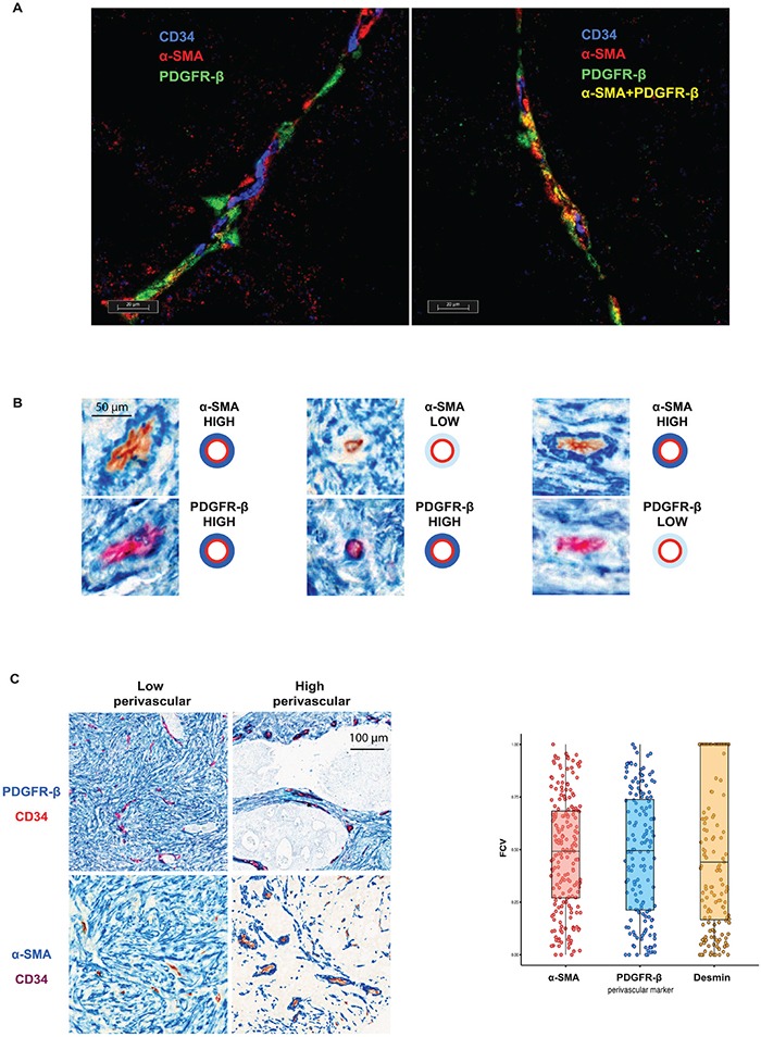
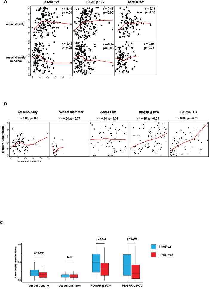
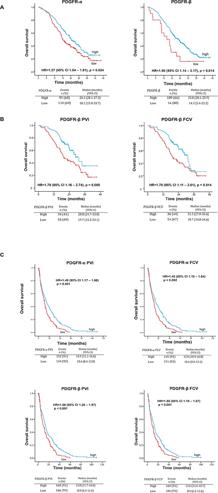
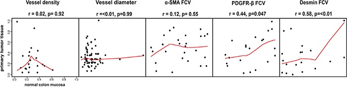
Perivascular cells (PC) were recently implied as regulators of metastasis and immune cell activity. Perivascular heterogeneity in clinical samples, and associations with other tumor features and outcome, remain largely unknown.Here we report a novel method for digital quantitative analyses of vessel characteristics and PC, which was applied to two collections of human metastatic colorectal cancer (mCRC).Initial analyses identified marker-defined subsets of PC, including cells expressing PDGFR-β or α-SMA or both markers. PC subsets were largely independently expressed in a manner unrelated to vessel density and size. Association studies implied specific oncogenic mutations in malignant cells as determinants of PC status. Semi-quantitative and digital-image-analyses-based scoring of the NORDIC-VII cohort identified significant associations between low expression of perivascular PDGFR-α and -β and shorter overall survival. Analyses of the SPCRC cohort confirmed these findings. Perivascular PDGFR-α and -β remained independent factors for survival in multivariate analyses.Overall, our study identified host vasculature and oncogenic status as determinants of tumor perivascular features. Perivascular PDGFR-α and -β were identified as novel independent markers predicting survival in mCRC. The novel methodology should be suitable for similar analyses in other tumor collections.

Keywords: PDGFR; cancer associated fibroblasts; colorectal cancer; perivascular cells; tumor stroma.


Conflict of interest statement

The authors declare no conflicts of interest.

Figures

Figure 1
Figure 1. A. Individual perivascular cells express different markers
The triple staining with both perivascular cell markers (PDGFR-β (red) or α-SMA (green)) and CD34 (blue) demonstrate presence of perivascular cells of three distinct perivascular cell types: PDGFR-β- and α-SMA-single positive cells and double-positive cells. B. Individual vessels are characterized by relatively independent perivascular expression of PDGFR-β and α-SMA. Serial sections of three vessels stained with α-SMA (blue staining, upper panels) or PDGFR-β (blue staining, lower panel) and CD34 (brown or red staining) showing independent perivascular expression of PDGFR-β and α-SMA. Note in left part an α-SMA-high / PDGFR-β-high vessel, in the middle part an α-SMA-low / PDGFR-β –high vessel and in right an α-SMA-high / PDGFR-β-low vessel. C. Inter-tumoral variation of perivascular marker expression. Left panel: Representative images of PDGFR-β and α-SMA (blue) double staining with CD34 (red or brown) showing areas of low or high perivascular marker expression. Note different intensity of perivascular staining (blue) in different cases. Right panel: Distribution of the fraction of covered vessels with PDGFR-β, α-SMA and Desmin in Nordic-VII cohort (right panel). The cases of the cohort are characterized by heterogeneous perivascular cell coverage defined by three markers.
Figure 2
Figure 2. A. Associations between perivascular status and vessel size or vessel density
Perivascular α-SMA, PDGFR-β and desmin status was largely independent of both vessel density and vessel size in analyses correlating these features on a case basis. Spearman rank test used for statistical analysis. B. Associations of vessel size, vessel density or perivascular status between primary tumor and normal colon mucosa. Perivascular PDGFR-β and desmin status in normal and tumor tissue showed significant correlations. Notably, vessel density, vessel size and perivascular α-SMA did not show this correlation. Horizontal axes show tumor tissue data, vertical axes show normal tissue data. Spearman rank test used for statistical analysis. C. Associations between perivascular and vascular status BRAF-mutation. Statistically significant differences were detected regarding perivascular PDGFR-α, PDGFR-β and vessel density in tumors with or without BRAF mutation. Average values of vessel number analyzed with regard to presence of correlation inside one tumor/peritumoral area. p values determined by Mann–Whitney U test.
Figure 3
Figure 3. A. Associations between perivascular expression of PDGFR-α and PDGFR-β and OS in the NORDIC-VII cohort (manual scoring)
Low expression of both PDGFR-α and PDGFR-β is associated with statistically significantly shorter OS in the total study population. B. Associations between perivascular metrics PVI and FCV of PDGFR-β and OS or PFS in the NORDIC-VII cohort. Low PVI and FCV of PDGFR-β are associated with statistically significantly shorter OS in the total study population. C. Associations between perivascular metrics PVI and FCV of PDGFR-α and PDGFR-β and OS in the SPCRC cohort. Low PVI and FCV of both PDGFR-α and PDGFR-β are associated with statistically significantly shorter OS in the total study population.
Figure 4
Figure 4. Associations of vessel size, vessel density or perivascular status between primary tumor and distant metastases
Perivascular PDGFR-β and desmin status in primary tumor tissue and in metastatic tissue showed significant correlations. Notably, vessel density, vessel size and perivascular α-SMA did not show this correlation. Horizontal axes show tumor tissue data, vertical axes show metastatic tissue data. Spearman rank test used for statistical analysis.

Similar articles

Cited by

References

    1. Armulik A, Genove G, Betsholtz C. Pericytes: Developmental, Physiological, and Pathological Perspectives, Problems, and Promises. Dev Cell. 2011;21:193–215. - PubMed
    1. Dulauroy S, Di Carlo SE, Langa F, Eberl G, Peduto L. Lineage tracing and genetic ablation of ADAM12(+) perivascular cells identify a major source of profibrotic cells during acute tissue injury. Nat Med. 2012;18:1262–1270. - PubMed
    1. Goritz C, Dias DO, Tomilin N, Barbacid M, Shupliakov O, Frisen J. A Pericyte Origin of Spinal Cord Scar Tissue. Science. 2011;333:238–242. - PubMed
    1. Lin SL, Kisseleva T, Brenner DA, Duffield JS. Pericytes and Perivascular Fibroblasts Are the Primary Source of Collagen-Producing Cells in Obstructive Fibrosis of the Kidney. Am J Pathol. 2008;173:1617–1627. - PMC - PubMed
    1. Humphreys BD, Lin SL, Kobayashi A, Hudson TE, Nowlin BT, Bonventre JV, Valerius MT, McMahon AP, Duffield JS. Fate Tracing Reveals the Pericyte and Not Epithelial Origin of Myofibroblasts in Kidney Fibrosis. Am J Pathol. 2010;176:85–97. - PMC - PubMed

Publication types

MeSH terms

Substances