. 2017 Mar;232(3):517-525.
doi: 10.1002/jcp.25445. Epub 2016 Jun 10.

Non-Metabolic Role of PKM2 in Regulation of the HIV-1 LTR

Affiliations

Non-Metabolic Role of PKM2 in Regulation of the HIV-1 LTR

Satarupa Sen et al. J Cell Physiol. 2017 Mar.

Abstract

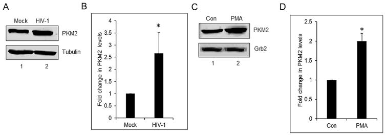
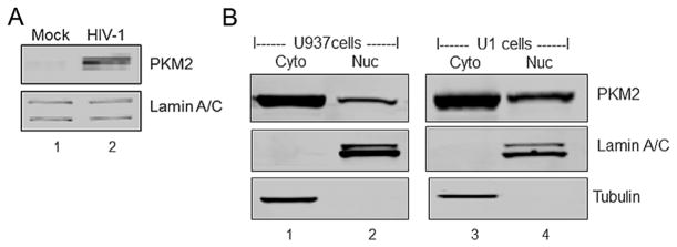
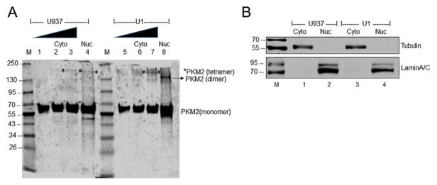
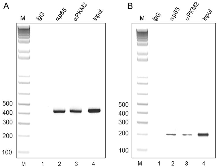
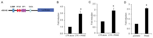
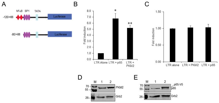
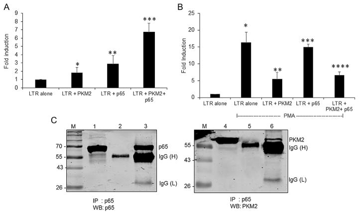
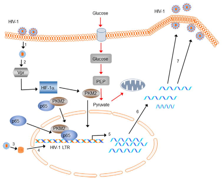
Identification of cellular proteins, in addition to already known transcription factors such as NF-κB, Sp1, C-EBPβ, NFAT, ATF/CREB, and LEF-1, which interact with the HIV-1 LTR, is critical in understanding the mechanism of HIV-1 replication in monocytes/macrophages. Our studies demonstrate upregulation of pyruvate kinase isoform M2 (PKM2) expression during HIV-1SF162 infection of monocyte/macrophages and reactivation of HIV-1 in U1 cells, a macrophage model of latency. We observed that HIV-1SF162 infection of monocyte/macrophages and reactivation of HIV-1 in U1 cells by PMA resulted in increased levels of nuclear PKM2 compared to PMA-induced U937 cells. Furthermore, there was a significant increase in the nuclear dimeric form of PKM2 in the PMA-induced U1 cells in comparison to PMA-induced U937 cells. We focused on understanding the potential role of PKM2 in HIV-1 LTR transactivation. Chromatin immunoprecipitation (ChIP) analysis in PMA-activated U1 and TZM-bl cells demonstrated the interaction of PKM2 with the HIV-1 LTR. Our studies show that overexpression of PKM2 results in transactivation of HIV-1 LTR-luciferase reporter in U937, U-87 MG, and TZM-bl cells. Using various truncated constructs of the HIV-1 LTR, we mapped the region spanning -120 bp to -80 bp to be essential for PKM2-mediated transactivation. This region contains the NF-κB binding site and deletion of this site attenuated PKM2-mediated activation of HIV-1 LTR. Immunoprecipitation experiments using U1 cell lysates demonstrated a physical interaction between PKM2 and the p65 subunit of NF-κB. These observations demonstrate for the first time that PKM2 is a transcriptional co-activator of HIV-1 LTR. J. Cell. Physiol. 232: 517-525, 2017. © 2016 Wiley Periodicals, Inc.


Figures

Fig. 1
Fig. 1
HIV-1 induces PKM2 expression in monocytes/macrophages and in PMA-induced U1 cells. (A) Western blot of PBMC lysates. Lane 1: mock infected (mock); lane 2: infected with HIV-1 SF162 (HIV-1) showing PKM2 and α-tubulin protein expression from whole cell lysates after infection for 6 days. (B) Densitometric analysis of PKM2 expression levels normalized to α-tubulin levels plotted as fold change in PKM2 levels. *P <0.05. (C) Western blot of undifferentiated U1 cell lysates. Lane 1: (Con) and lane 2: PMA-treated U1 cells (PMA) showing PKM2 and Grb2 protein expression in cell lysates after 72 h of differentiation. (D) Densitometric analysis of PKM2 expression levels normalized to Grb2 levels, plotted as fold change in PKM2 levels. *P <0.05.
Fig. 2
Fig. 2
HIV-1 induces nuclear translocation of PKM2 in monocytes/macrophages and PMA induced U937 and U1 cells. (A) Western blot of PBMC lysates. Lane 1: mock infected (mock). Lane 2: infected with HIV-1 SF162 (HIV-1) showing levels of PKM2 and lamin A/C protein in nuclear fractions obtained from cells after infection for 6 days. (B) Western blot of cytoplasmic fraction and nuclear fractions. Lane 1: cytoplasmic fraction (Cyto) and lane 2: nuclear fraction (Nuc) of U937 cells differentiated with PMA. Lane 3: cytoplasmic fraction (Cyto) and lane 4: nuclear fraction (Nuc) of U1 cells differentiated with PMA. Western blots showing levels of PKM2, tubulin in cytoplasmic fraction and lamin A/C protein in nuclear fractions.
Fig. 3
Fig. 3
Oligomerization status of PKM2 in cytoplasmic and nuclear fractions of PMA differentiated U937 and U1 cells. (A) Lane 1: 1 μg of cytoplasmic extract from U937 cells; lane 2: 2.5 μg of cytoplasmic extract from U937 cells; lane 3: 5 μg of cytoplasmic extract from U937 cells; lane 4: 40 μg of nuclear extract from U937 cells; Lane 5: 1 μg of cytoplasmic extract from U1 cells; lane 6: 2.5 μg of cytoplasmic extract from U1 cells; lane 7: 5 μg of cytoplasmic extract from U1 cells; lane 8: 40 μg of nuclear extract from U1 cells. *Tetrameric form of PKM2. Lane M: Molecular weight marker. (B) Western blot analysis of the cytoplasmic (lane 1) and nuclear fractions (lane 2) isolated from PMA induced U937, and cytoplasmic (lane 3) and nuclear fractions (lane 4) isolated from PMA induced U1 cells used in panel A to demonstrate the purity of fractionation. Tubulin was used as cytoplasmic marker and lamin A/C as a nuclear marker.
Fig. 4
Fig. 4
(A) The interaction of PKM2 and p65 in vivo with HIV-1 LTR in U1 cells induced with PMA was analyzed using ChIP analysis. DNA isolated from chromatin immunoprecipitated with Lane 1: normal rabbit IgG (negative control, IgG); Lane 2: p65 antibodies (p65), and lane 3: PKM2 antibodies (PKM2) was amplified by PCR using primers described in the materials and methods. Lane 4: Total chromatin used as the input (input) and amplified by PCR. Lane: M: DNA size ladder (B). The interaction of PKM2 and p65 in vivo with HIV-1 LTR in TZM-bl cells induced with PMA was analyzed using ChIP analysis. DNA isolated from chromatin immunoprecipitated with Lane 1: normal rabbit IgG (negative control, IgG); Lane 2: p65 antibodies (p65) and lane 3: PKM2 antibodies (PKM2) was amplified by PCR using primers described in the materials and methods. Lane 4: Total chromatin used as the input (lane: input) and amplified by PCR. Lane: M: DNA size ladder.
Fig. 5
Fig. 5
(A) Schematic representation of the HIV-1 LTR/LUC construct. The −456/+66 construct harbors the C/EBP site, two NF-κB, three SP1 sites, and the TATAA box. (B) Fold induction of HIV-1 LTR (−456/+66) promoter activity in U937 cells transiently cotransfected with 0.3 μg of HIV-1 LTR/Luc reporter vector and 0.6 μg of an empty pcDNA3 expression vector (LTR alone), and 0.3 μg of HIV-1 LTR/Luc reporter vector and 0.6 μg expression vector for PKM2 (LTR + PKM2). RLU/s was normalized to protein concentration and expressed as fold induction wherein the activity in HIV-1 LTR/Luc alone transfected cells is set at 1. Statistical significance (*P <0.007 between LTR alone and LTR + PKM2) was analyzed by Student’s t-test. (B) Fold induction of HIV-1 LTR (−456/+66) promoter activity in U-87 MG cells transiently cotransfected with 0.25 μg of HIV-1 LTR/Luc reporter vector and 0.25 μg of an empty expression vector (LTR alone), and 0.25 μg of HIV-1 LTR/LUC and 0.25 μg expression vector for PKM2 (LTR + PKM2). RLU/s was normalized to protein concentration and expressed as fold induction wherein the activity in HIV-1 LTR/Luc alone transfected cells is set at 1. Statistical significance (*P <0.001 between LTR alone and LTR + PKM2) was analyzed by Student’s t-test. (C) Transient transfection of TZM-bl cells harboring integrated copies of HIV-1 LTR/LUC. pcDNA represents cells transfected with 0.25 μg of pcDNA3 empty vector and PKM2 represents cells transfected with 0.25 μg of PKM2 expression vector. RLU/s was normalized to protein concentration and expressed as fold induction wherein the activity in pcDNA3 alone-transfected cells is set at 1. Statistical significance (*P <0.002 between pcDNA3 and PKM2) was analyzed by Student’s t-test.
Fig. 6
Fig. 6
(A) Schematic representation of the HIV-1 LTR/LUC deletion constructs. The −120/+66 construct harbors two NF-κB, three SP1 sites, and TATAA box. The −80/+66 construct harbors only three SP1 sites and TATAA box. (B) Fold induction of HIV-1 LTR (P−120/+66) promoter activity in U-87 MG cells transiently cotransfected with 0.25 μg HIV-1 LTR/Luc reporter vector and 0.25 μg of an empty expression vector (LTR alone), or an expression vector for p65 (LTR + p65) or PKM2 (LTR + PKM2). RLU/s was normalized to protein concentration and expressed as fold induction wherein the activity in HIV-1 LTR/Luc alone transfected cells is set at 1. Statistical significance (*P <0.002 between LTR alone and LTR + p65 transfected cells; **P <0.003 between LTR alone and LTR + PKM2 transfected cells) was analyzed by Student’s t-test. (C) Fold induction of HIV-1 LTR (−88/+66) promoter activity in U-87 MG cells transiently cotransfected with 0.25 μg HIV-1 LTR/Luc reporter vector and 0.25 μg of an empty expression vector (LTR alone) or 0.25 μg of an expression vector for PKM2 (LTR + PKM2) or p65 (LTR + p65). Cells were harvested 48 h post-transfection to measure luciferase activity. RLU/s was normalized to protein concentration and expressed as fold induction wherein the activity in HIV-1 LTR/Luc alone transfected cells is set at 1. (D) Representative Western blot analysis of expression levels of PKM2 in U-87 MG cells. Lane 1: pcDNA 3 empty vector; lane 2: PKM2 cDNA vector. (E) Representative Western blot analysis of expression levels of p65 in U-87 MG cells. Lane 1: pcDNA empty vector; lane 2: p65 cDNA vector. p65 denotes the endogenous p65 protein and p65-V5 represents the V5-tagged p65 protein.
Fig. 7
Fig. 7
(A) Synergistic activation of HIV-1 LTR by p65 and PKM2. Fold induction of HIV-1 LTR (−120/+66) promoter activity in U-87 MG cells transiently cotransfected with 0.25 μg of HIV-1 LTR/Luc reporter vector and 0.5 μg of an empty expression vector (LTR alone), or 0.25 μg of an expression vector for p65 (LTR + p65), or 0.25 μg of PKM2 (LTR + p65), and 0.25 μg of p65 and PKM2 expression vectors (LTR + p65 + PKM2). Cells were harvested 48 h post transfection to measure luciferase activity. RLU/s was normalized to protein concentration and expressed as fold induction wherein the activity in HIV-1 LTR/Luc alone transfected cells is set at 1. Statistical significance (*P <0.034 between LTR alone and LTR + PKM2 transfected cells; **P <0.016 between LTR alone and LTR+ p65; ***P <0.0003 between LTR alone and PKM2 + p65 transfected cells) was analyzed by Student’s t-test. (B) Fold induction of HIV-1 LTR (−120/+66) promoter activity in U-87 MG cells transiently cotransfected with 0.25 μg of HIV-1 LTR/Luc reporter vector and 0.5 μg of an empty expression vector (LTR alone), or 0.25 μg of an expression vector for p65 (LTR + p65), or 0.25 μg of PKM2 (LTR + p65), and 0.25 μg of p65 and PKM2 expression vectors (LTR + p65 + PKM2) and then treated with PMA for 24 h post transfection. Cells were harvested 48 h post-transfection to measure luciferase activity. RLU/s was normalized to protein concentration and expressed as fold induction wherein the activity in HIV-1 LTR/Luc alone transfected cells is set at 1. Statistical significance (*P <0.0004 between LTR alone and LTR alone transfected cells treated with PMA; **P <0.008 between LTR alone and LTR + PKM2 transfected cells treated with PMA; ***P <0.00005 between LTR alone and LTR + p65 transfected cells treated with PMA; ****P <0.003 between LTR alone and LTR+ p65 and PKM2 transfected cells treated with PMA) was analyzed by Student’s t-test. (C) PKM2 interacts with NF-κB p65 in U1 cells. Left panel: IP-Western analysis of whole cell lysates from PMA induced U1 cells immunoprecipitated with p65 antibodies or isotype specific antibodies and immunoblotted with p65 antibodies. Lane 1: whole cell lysate, lane 2: cell lysates IP with isotype specific antibody, and lane 3: cell lysates IP with p65 antibodies. Right panel: IP-Western analysis of whole cell lysates from PMA induced U1 cells immunoprecipitated with p65 antibodies or isotype specific antibodies and immunoblotted with PKM2 antibodies. Lane 1: whole cell lysate, lane 2: cell lysates IP with isotype specific antibody, and lane 3: cell lysates IP with p65 antibodies. IgG(H): heavy chain, IgG(L): light chain.
Fig. 8
Fig. 8
Schematic representation of the non-metabolic role of PKM2 in HIV-1-infected macrophages. HIV-1 infection of monocyte/macrophages and virus entry and core release (step 1). Virion-associated Vpr (step 2) in an HIF-1α-dependent manner induces PKM2 (Barrero et al., 2013). Following reverse transcription, proviral DNA (step 3) is imported into the nucleus and integrated into the host genome (step 4). Viral infection or reactivation of integrated virus (latent infection) results in the induction and nuclear translocation of PKM2 and p65 independently from the cytoplasm or by forming a functional complex (p65/PKM2) in the cytoplasm and subsequent nuclear translocation. p65/PKM2 interacts with the kB sites on HIV-1 LTR and synergistically activates transcription (step 5). Fully spliced mRNAs and partially spliced and unspliced RNAs are then transported to the cytoplasm and translated (step 6). Unspliced RNA is transported to the cell membrane, where Env, Gag, and Gag-Pol polyproteins assemble and incorporates into new virus particles (step 7). The new virus particle is then released after maturation.

Similar articles

Cited by

References

    1. Ahmad N, Venkatesan S. Nef protein of HIV-1 is a transcriptional repressor of HIV-1 LTR. Science. 1988;241:1481–1485. - PubMed
    1. Amini S, Clavo A, Nadraga Y, Giordano A, Khalili K, Sawaya BE. Interplay between cdk9 and NF-kappaB factors determines the level of HIV-1 gene transcription in astrocytic cells. Oncogene. 2002;21:5797–5803. - PubMed
    1. Anastasiou D, Yu Y, Israelsen WJ, Jiang JK, Boxer MB, Hong BS, Tempel W, Dimov S, Shen M, Jha A, Yang H, Mattaini KR, Metallo CM, Fiske BP, Courtney KD, Malstrom S, Khan TM, Kung C, Skoumbourdis AP, Veith H, Southall N, Walsh MJ, Brimacombe KR, Leister W, Lunt SY, Johnson ZR, Yen KE, Kunii K, Davidson SM, Christofk HR, Austin CP, Inglese J, Harris MH, Asara JM, Stephanopoulos G, Salituro FG, Jin S, Dang L, Auld DS, Park HW, Cantley LC, Thomas CJ, Vander Heiden MG. Pyruvate kinase M2 activators promote tetramer formation and suppress tumorigenesis. Nat Chem Biol. 2012;8:839–847. - PMC - PubMed
    1. Barrero CA, Datta PK, Sen S, Deshmane S, Amini S, Khalili K, Merali S. HIV-1 Vpr modulates macrophage metabolic pathways: A SILAC-based quantitative analysis. PLoS ONE. 2013;8:e68376. - PMC - PubMed
    1. Brass AL, Dykxhoorn DM, Benita Y, Yan N, Engelman A, Xavier RJ, Lieberman J, Elledge SJ. Identification of host proteins required for HIV infection through a functional genomic screen. Science. 2008;319:921–926. - PubMed

MeSH terms

LinkOut - more resources