. 2016 Jun 1;11(6):e0155422.
doi: 10.1371/journal.pone.0155422. eCollection 2016.

APOBEC4 Enhances the Replication of HIV-1

Affiliations

APOBEC4 Enhances the Replication of HIV-1

Daniela Marino et al. PLoS One. .

Abstract

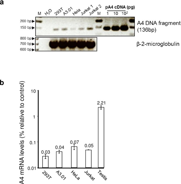
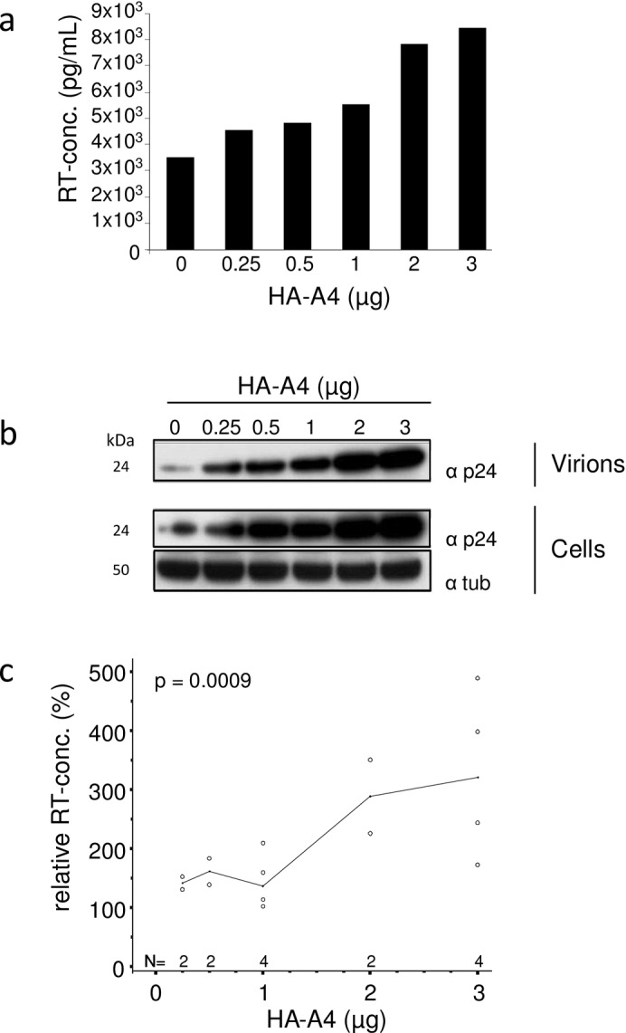
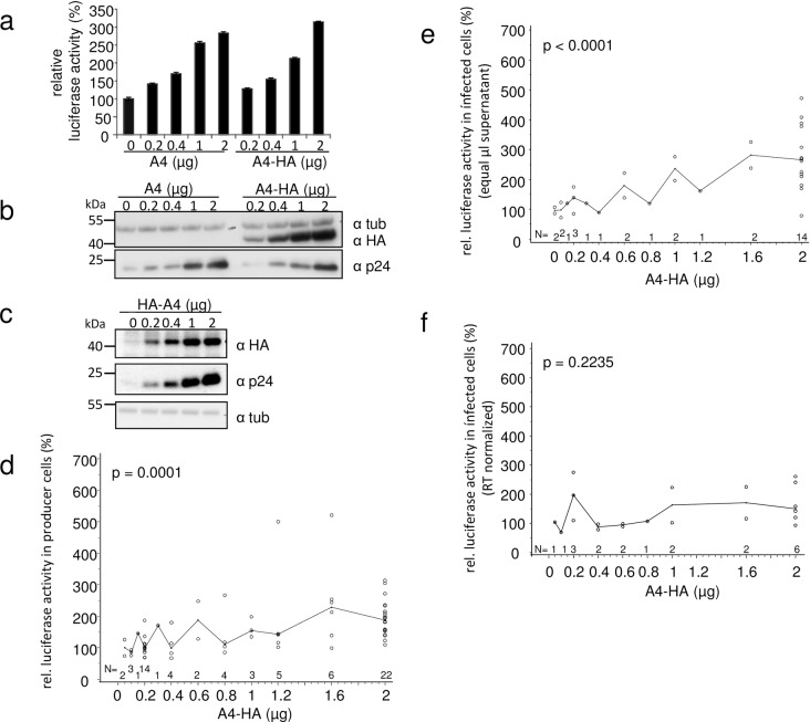
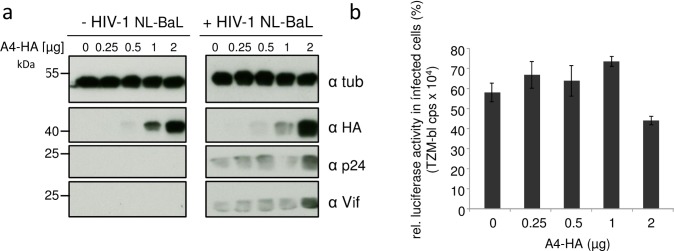
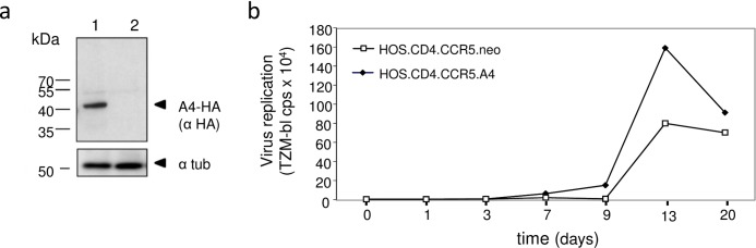
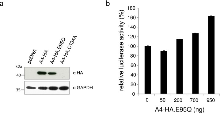
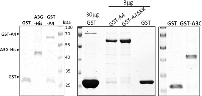
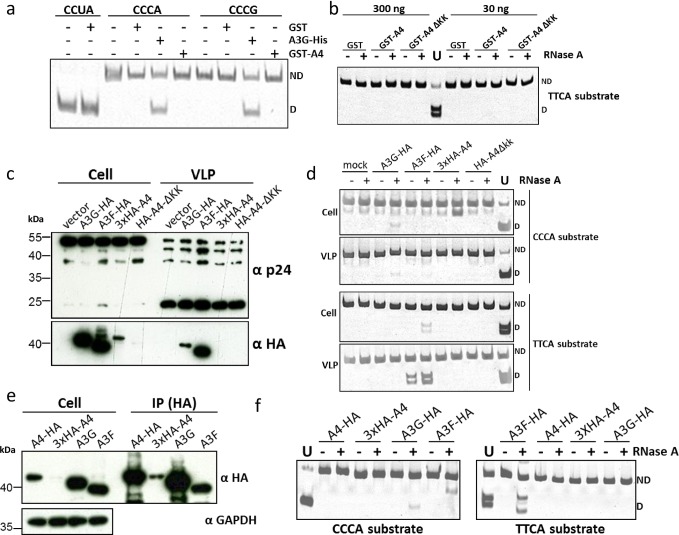
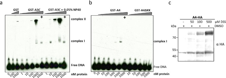
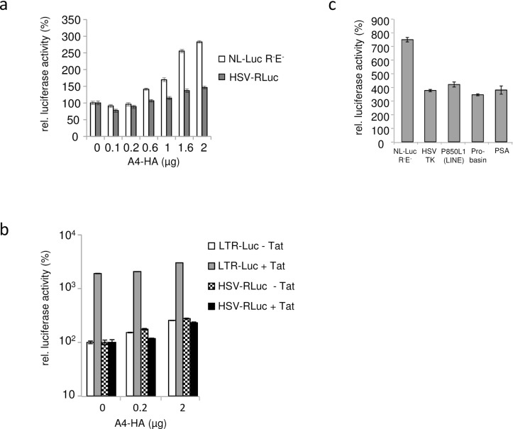
APOBEC4 (A4) is a member of the AID/APOBEC family of cytidine deaminases. In this study we found a high mRNA expression of A4 in human testis. In contrast, there were only low levels of A4 mRNA detectable in 293T, HeLa, Jurkat or A3.01 cells. Ectopic expression of A4 in HeLa cells resulted in mostly cytoplasmic localization of the protein. To test whether A4 has antiviral activity similar to that of proteins of the APOBEC3 (A3) subfamily, A4 was co-expressed in 293T cells with wild type HIV-1 and HIV-1 luciferase reporter viruses. We found that A4 did not inhibit the replication of HIV-1 but instead enhanced the production of HIV-1 in a dose-dependent manner and seemed to act on the viral LTR. A4 did not show detectable cytidine deamination activity in vitro and weakly interacted with single-stranded DNA. The presence of A4 in virus producer cells enhanced HIV-1 replication by transiently transfected A4 or stably expressed A4 in HIV-susceptible cells. APOBEC4 was capable of similarly enhancing transcription from a broad spectrum of promoters, regardless of whether they were viral or mammalian. We hypothesize that A4 may have a natural role in modulating host promoters or endogenous LTR promoters.


Conflict of interest statement

Competing Interests: The authors have declared that no competing interests exist.

Figures

Fig 1
Fig 1. Differential expression of A4.
(a) A4 expression was determined by semi-quantitative RT-PCR. Low level A4 amplification by PCR using equal amount of cDNA prepared from total RNA of 293T, A3.01, Hela, and Jurkat cell lines. As a control, β-2-microglobulin (ß-2-M) cDNA was amplified. Water instead of template served as a background control and a plasmid coding for A4 cDNA (pA4 cDNA) served as a positive control. M: 50 bp DNA ladder. (b) Levels of A4 expression were determined by quantitative real-time RT-PCR and measured relative to endogenous HPRT1 RNA levels. A4 is expressed at a high level in human testis tissue, while 293T, HeLa, A3.01 and Jurkat cells exhibit very low A4 expression. Error bars indicate standard deviation.
Fig 2
Fig 2. Expression of the A4-HA fusion proteins.
(a) Schematic representation of protein domains and motifs found in the human A4 protein and tested variants. Zn2+: presumed zinc-binding domain. HA (white boxes): HA-tag. KKKKKGKK: polylysine domain. (b) Increasing amounts of A4-HA (3xHA-tags), A3G-HA (1xHA-tag) and A3A-HA (3xHA-tags) expression plasmids were transfected into 293T cells followed by immunoblot analysis of the transfected cells using an anti-HA antibody. Immunoblot analysis with anti-tubulin (tub) antibody served as loading control. α, anti.
Fig 3
Fig 3. Subcellular localization of A4 in transfected cells.
Immunofluorescence confocal laser scanning microscopy images of HeLa cells transfected with N- or C-terminal HA-tagged A4 (HA-A4 and A4-HA). (a, b) HA-A4 proteins show cytoplasmic and nuclear localization. (c, d) A4-HA proteins show cytoplasmic localization. (a, c, e, f) x-y optical sections. (b, d) x-z vertical scanning image of indicated cells (see asterisks). (e) Mock transfected cells (no A4). (f) HA-A4ΔKK transfected cells show cytoplasmic and nuclear localization. To detect A4 (green) immunofluorescence, cells were stained with an anti-HA antibody. Nuclei (blue) were visualized by DAPI staining. α, anti.
Fig 4
Fig 4. A4 enhances the expression of HIV-1.
(a) HIV-1 genome expression plasmid was co-transfected with increasing amounts of HA-A4 expression plasmid, as indicated. A4 increases the production of HIV-1 particles as measured by the RT activity in the supernatant of the transfected cells. (b) Immunoblot analysis of virions and transfected 293T cells (same cells as in (a)). Immunoblots of virions and cell lysates were probed with anti-p24 (capsid) antibody. Anti-tubulin (tub) antibody served as loading control. α, anti. (c) RT concentrations in the supernatant of cells co-transfected with HA-A4 and HIV-1 plasmids relative to supernatant of cells co-transfected with empty vector and HIV-1, as in (a), summary of four independent experiments, median indicated. Evaluation of RT activity data was performed by means of a multifactorial analysis of variance (ANOVA).
Fig 5
Fig 5. Presence of A4 does not affect HIV-1 infectivity.
HIV-1 reporter virus NL-Luc R-E- (VSV-G) was produced in 293T cells in the presence of increasing amounts of A4 (no tag) and A4-HA (C-terminal HA-tag). A4 and A4-HA increase in a dose-dependent manner both (a) the virus-encoded luciferase activity and (b) the expression of intracellular viral capsid (p24) in the transfected virus producing cells as demonstrated by immunoblot analysis (same cell lysates used in (a) and (b)). Error bars indicate standard deviation. (c) Immunoblot analysis of intracellular viral p24 (capsid) expression. Similar as in (a) and (b), NL-Luc R-E-/VSV-G was co-transfected with increasing amounts of HA-A4 plasmid (N-terminal HA-tag), as indicated. Immunoblots of cells were probed with anti-p24 (capsid) antibody. A4-HA expression in transfected cells was detected by immunoblotting using anti-HA antibody. Anti-tubulin (tub) antibody served as loading control. α, anti. (d) Relative viral luciferase activity in cells co-transfected with A4-HA and HIV-1 plasmids, as in (a). Summary of 28 independent experiments, median indicated. A4-HA was transfected in increasing amounts. (e) Equal volumes of supernatants of cells co-transfected with NL-Luc R-E-/VSV-G and increasing amounts of A4-HA were used to infect HOS cells. Intracellular luciferase activities were determined in infected cells; summary of 16 experiments (a subset of the experiments shown in (d)), median is indicated. (f) A subset of samples (seven experiments) used in (e) was quantified for RT concentrations. RT normalized supernatants of cells co-transfected with NL-Luc R-E-/VSV-G and increasing amounts of A4-HA were used to infect HOS cells. Intracellular luciferase activities determined in infected cells, median is indicated. (d—f) Statistical evaluation of reporter luciferase activity data was performed by means of a multifactorial ANOVA.
Fig 6
Fig 6. A4 enhances expression of CCR5-tropic HIV-1.
(a) Increasing amounts of A4-HA expression plasmid were co-transfected with HIV-1 NL-BaL and immunoblot analysis of co-transfected 293T cells were performed. Immunoblots were probed with anti-p24 (capsid), anti-Vif, anti-HA and anti-tubulin (tub) antibodies. α, anti. (b) Infectivity of RT-normalized viral supernatant of the transfected cells from (a) were used to infect TZM-bl luciferase reporter cells. cps, counts per second. Data are represented as the mean with SD. Statistically significant differences between no A4 and A4 groups were analyzed using the unpaired Student’s t-test with GraphPad Prism version 5 (GraphPad software, San Diego, CA, USA). Validity of the null hypothesis was verified with significance level at α value = 0.05. NS: not significant.
Fig 7
Fig 7. A4 enhances multiple cycle replication of HIV-1.
(a) Immunoblot analysis of A4-HA expressing HOS.CD4.CCR5.A4 cells (1) and empty retroviral vector just encoding G418-resistance containing HOS.CD4.CCR5.neo cells (2) using an anti-HA antibody. Cell lysates were also analyzed for equal amounts of total proteins by using anti-tubulin antibody. (b) HOS.CD4.CCR5.A4 and HOS.CD4.CCR5.neo cells were infected with HIV-1 clone NL-BaL, MOI of 0.01. Virus replication was monitored by testing the cell supernatants on TZM-bl cells and measuring luciferase activity.
Fig 8
Fig 8. Active site mutation has no influence on A4 activity.
(a) Protein expression of A4-HA, A4-HA.E95Q and A4-HA.C134A detected by anti-HA immuno blot analysis, showing equal amounts of A4-HA and A4-HA.E95Q, but lack of A4-HA.C134A expression in transfected cells. (b) HIV-1 reporter virus (NL-Luc R-E-) was co-transfected with increasing amounts of expression plasmid for A4-HA.E95Q. Virus encoded luciferase activity in the transfected cells was enhanced by A4-HA.E95Q in a dose-dependent manner.
Fig 9
Fig 9. Recombinantly produced and affinity purified E. coli-derived GST, GST-A3C, GST-A4, GST-A4-ΔKK proteins and 293T cell-derived A3G-His protein were resolved on a 10% SDS gel.
Purity of the proteins was determined by staining the gel with Coomassie blue. GST-A4, A3G-His and GST proteins are indicated according to their molecular mass.
Fig 10
Fig 10. A4 does not deaminate single stranded DNA.
(a) Deamination activity of A4 was tested on two different oligonucleotide substrates containing nucleotide sequences CCCA or CCCG. The A3G-His fusion protein was incubated with CCCA and CCCG containing substrates and served as positive control for deamination resulting in 40-bp DNA fragments. Oligonucleotide CCUA served as a marker to denote the deaminated product after Eco147I cleavage; ND: not deaminated; D: deaminated. (b) Deamination experiment using TTCA containing oligonucleotide and GST-purified A4 proteins, RNAse A treatment was included; ND: not deaminated; D: deaminated. (c) Immuno blot analysis of cell lysates and virus lysate of A3G-HA, A3F-HA, 3xHA-A4 and HA-A4-ΔKK expressing cells and HIV virus like particles (VLP), respectively. Anti-HA staining indicates the presence of HA-tagged A3 and A4 proteins, while anti-p24 antibody detects HIV-1 capsid proteins. (d) Deamination assay using transfected 293T cell lysate (from experiment shown in (c)). RNAse A treatment was included; ND: not deaminated; D: deaminated. (e) Immuno blot analysis of cell lysate and immunoprecipitate (IP) fraction of A3 and A4 proteins. (f) Deamination assay using the immunoprecipitated APOBEC proteins (from experiment shown in (e)). RNAse A treatment was included; ND: not deaminated; D: deaminated.
Fig 11
Fig 11. A4 interacts weakly with single-stranded DNA.
EMSA with purified, GST-A3C (a), GST-A4 and GST-A4ΔKK (b) performed with 30 nt single stranded target DNA labeled with 3’-labeled with biotin. Indicated amounts of protein (at the bottom of blot) were titrated with 10 nM of DNA. (+) indicates presence of competitor DNA, which is unlabeled 80 nt DNA (200-fold molar excess), as used for deamination assay to demonstrate specific binding of protein to DNA being causative for the shift. For GST-A3C (a) a separate panel was added for reactions containing 0.05% NP-40 detergent. (c) A4-HA crosslinking by DSS. DSS was added to the cleared cell lysates to reach the indicated DSS concentrations. The blot was probed with anti HA antibody to detect monomeric and dimeric forms of A4-HA.
Fig 12
Fig 12. A4 enhances expression of luciferase reporter genes driven by various viral and cellular promoters.
(a) Dual luciferase reporter assay was performed two days post co-transfection of NL-Luc R-E- and HSV-TK promoter Renilla luciferase (HSV-RLuc) with and without A4-HA, relative luciferase activities are shown. (0) indicates transfections in the absence of A4-HA plasmid. A4-HA was transfected in increasing amounts. (b) Relative luciferase activities after co-transfection of LTR-Luc (LTR of HIV-1 driving firefly luciferase) with A4-HA or HSV-TK promoter Renilla luciferase (HSV-RLuc) with A4-HA and with and without Tat expression plasmid (c) Luciferase activities driven by various viral (LTR, HSV TK) or cellular promoters (LINE, Probasin, PSA) in presence of the transfected A4-HA expression plasmid, relative to luciferase activity in cells without A4 expression. Total amounts of luciferase expression plasmid and total plasmid DNA was kept constant within all experiments. Error bars indicate standard deviation.

References

    1. Jarmuz A, Chester A, Bayliss J, Gisbourne J, Dunham I, Scott J, et al. An anthropoid-specific locus of orphan C to U RNA-editing enzymes on chromosome 22. Genomics. 2002;79(3):285–96. - PubMed
    1. Navaratnam N, Sarwar R. An overview of cytidine deaminases. International journal of hematology. 2006;83(3):195–200. 10.1532/IJH97.06032 . - DOI - PubMed
    1. Rogozin IB, Basu MK, Jordan IK, Pavlov YI, Koonin EV. APOBEC4, a new member of the AID/APOBEC family of polynucleotide (deoxy)cytidine deaminases predicted by computational analysis. Cell Cycle. 2005;4(9):1281–5. . - PubMed
    1. Prohaska KM, Bennett RP, Salter JD, Smith HC. The multifaceted roles of RNA binding in APOBEC cytidine deaminase functions. Wiley interdisciplinary reviews RNA. 2014;5(4):493–508. 10.1002/wrna.1226 - DOI - PMC - PubMed
    1. Münk C, Willemsen A, Bravo IG. An ancient history of gene duplications, fusions and losses in the evolution of APOBEC3 mutators in mammals. BMC evolutionary biology. 2012;12:71 10.1186/1471-2148-12-71 - DOI - PMC - PubMed

LinkOut - more resources