. 2016 Jun 1:6:27280.
doi: 10.1038/srep27280.

HIF-1α and HIF-2α induced angiogenesis in gastrointestinal vascular malformation and reversed by thalidomide

Affiliations

HIF-1α and HIF-2α induced angiogenesis in gastrointestinal vascular malformation and reversed by thalidomide

Nan Feng et al. Sci Rep. .

Abstract

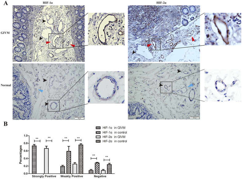
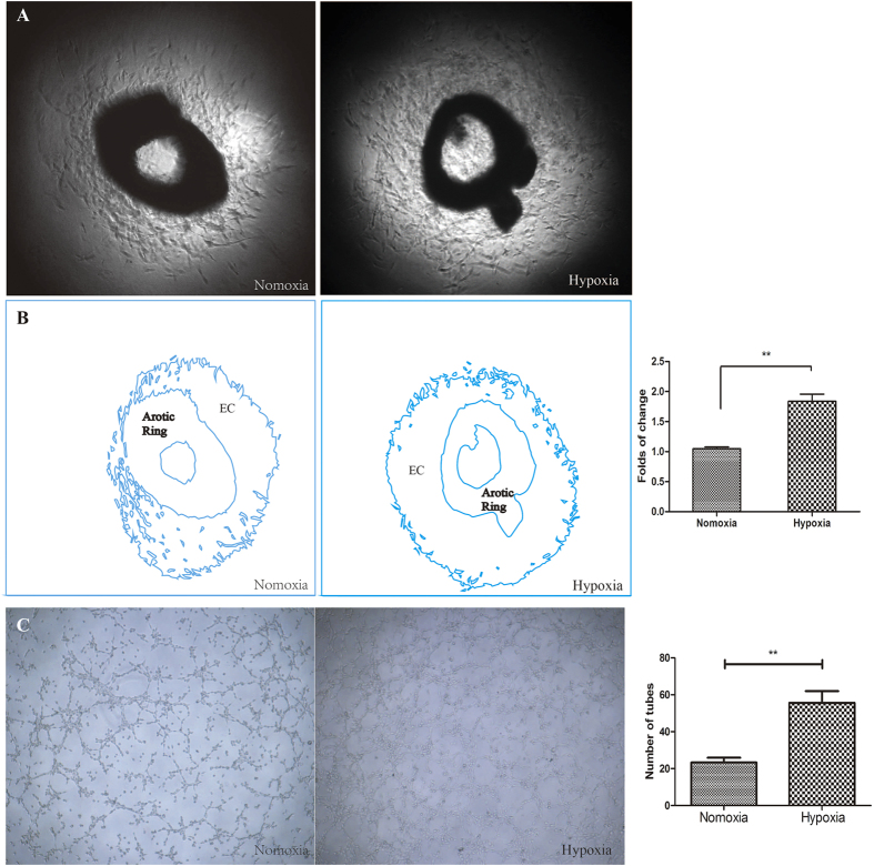
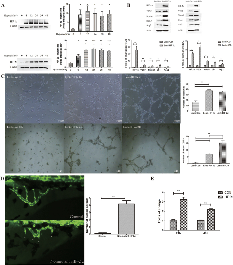
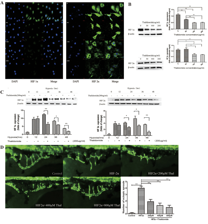
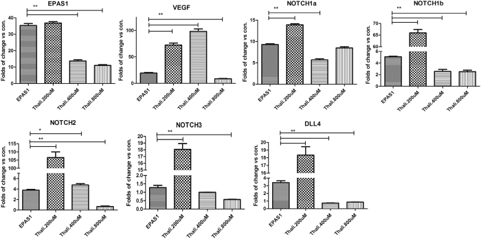
Thalidomide is used in clinical practice to treat gastrointestinal vascular malformation (GIVM), but the pathogenesis of GIVM is not clear. Hypoxia inducible factor 1 alpha (HIF-1α) and 2 alpha (HIF-2α/EPAS1) are in the same family and act as master regulators of the adaptive response to hypoxia. HIF-1α and HIF-2α are up-regulated in vascular malformations in intestinal tissues from GIVM patients, but not in adjacent normal vessels. Therefore, we investigated the role of HIF-1α and HIF-2α during angiogenesis and the mechanism of thalidomide action. In vitro experiments confirmed that vascular endothelial growth factor (VEGF) was a direct target of HIF-2α and that HIF-1α and HIF-2α regulated NOTCH1, Ang2, and DLL4, which enhanced vessel-forming of endothelial cells. Thalidomide down-regulated the expression of HIF-1α and HIF-2α and inhibited angiogenesis. In vivo zebrafish experiments suggested that HIF-2α overexpression was associated with abnormal subintestinal vascular (SIV) sprouting, which was reversed by thalidomide. This result indicated that thalidomide regulated angiogenesis via the inhibition of HIF-1α and HIF-2α expression, which further regulated downstream factors, including VEGF, NOTCH1, DLL4, and Ang2. The abnormally high expression of HIF-1α and HIF-2α may contribute to GIVM.


Figures

Figure 1
Figure 1
(A) The expression of HIF-1α and HIF-2α in gastrointestinal vascular malformations and normal vessels. Red arrow: strongly positive; Black arrow: weakly positive; Blue arrow: negative. (B) Percentages of positive and negative vessels in GIVM and normal tissues. **P < 0.01.
Figure 2
Figure 2. The effect of hypoxia on angiogenesis was assessed using mouse thoracic aortas and HUVECs.
(A) Aortic ring assays revealed increased EC proliferation under hypoxia. (B) The outline in (A) was drawn using Adobe Illustrator software, and the area was calculated. (C) Representative images of tube formation under normoxia and hypoxia. Tube formation was enhanced under hypoxia for 6 h. (EC: Endothelial Cells) **P < 0.01 vs. normoxia.
Figure 3
Figure 3
(A) Western blot determinations of HIF-1α and HIF-2α expression at different time points of hypoxia. *P < 0.05, **P < 0.01 vs. 0 hour. (B) The effect of HIF-1α and HIF-2α overexpression on the expression of VEGF, Notch1, DLL4, and Ang2. Western blot and RT-PCR demonstrated that HIF-1α and HIF-2α overexpression increased the expression of VEGF, Notch1, DLL4, and Ang2 protein and mRNA. *P < 0.05, **P < 0.01 vs. control. (C) Influence of HIF-1α and HIF-2α overexpression on angiogenesis 6 and 24 h after transfection of Lenti-HIF-1α and Lenti-HIF2α. Tube formation was enhanced 6 and 24 h after transfection. **P < 0.01 vs. control. (D) Fluorescence microscope observations of subintestinal vein sprouting in normal and HIF-2α-overexpressing zebrafish. *Indicates subintestinal vascular sprouts. HIF-2α overexpression significantly increased the number of subintestinal vascular sprouts. **P < 0.01 vs. control plasmid. (E) Dual luciferase reporter gene assay demonstrated that HIF-2α enhanced VEGF promoter activity. **P < 0.01 vs. control plasmid.
Figure 4
Figure 4. The inhibitory effect of thalidomide.
(A) Immunofluorescence indicated that HIF-1α and HIF-2α expression was down-regulated by thalidomide at different concentrations. (B) Western blots demonstrated that the expression of HIF-1α and HIF-2α decreased with 100 and 200 μg/ml of thalidomide. *P < 0.05, **P < 0.01. (C) Western blots demonstrated that thalidomide at 200 μg/ml inhibited the expression of HIF-1α and HIF-2α in HUVECs after hypoxic treatment for 24, 36, and 48 h. *P < 0.05, **P < 0.01. (D) Fluorescence microscope observations of the effect of thalidomide at different concentrations on vascular development in zebrafish with HIF-2α overexpression. **P < 0.01 vs. HIF-2α.
Figure 5
Figure 5. The expression of HIF-2α (EPAS1), VEGF, Notch1a, Notch1b, Notch2, Notch3, and DLL4 in the zebrafish model.
*P < 0.05, **P < 0.01.

References

    1. Regula J., Wronska E. & Pachlewski J. Vascular lesions of the gastrointestinal tract. Best Pract Res Cl Ga 22, 313–328, doi: 10.1016/j.bpg.2007.10.026 (2008). - DOI - PubMed
    1. Junquera F., Saperas E., de Torres I., Vidal M. T. & Malagelada J. R. Increased expression of angiogenic factors in human colonic angiodysplasia. Am J Gastroenterol 94, 1070–1076, doi: 10.1111/j.1572-0241.1999.01017.x (1999). - DOI - PubMed
    1. Li J. L. & Harris A. L. Crosstalk of VEGF and Notch pathways in tumour angiogenesis: therapeutic implications. Front Biosci (Landmark Ed) 14, 3094–3110 (2009). - PubMed
    1. Gaur P., Bose D., Samuel S. & Ellis L. M. Targeting tumor angiogenesis. Semin Oncol 36, S12–19, doi: 10.1053/j.seminoncol.2009.02.002 (2009). - DOI - PubMed
    1. Handra-Luca A. & Montgomery E. Vascular malformations and hemangiolymphangiomas of the gastrointestinal tract: morphological features and clinical impact. Int J Clin Exp Pathol 4, 430–443 (2011). - PMC - PubMed

Publication types

MeSH terms

Substances

LinkOut - more resources