. 2016 Jun 2:6:27144.
doi: 10.1038/srep27144.

The CebE/MsiK Transporter is a Doorway to the Cello-oligosaccharide-mediated Induction of Streptomyces scabies Pathogenicity

Affiliations

The CebE/MsiK Transporter is a Doorway to the Cello-oligosaccharide-mediated Induction of Streptomyces scabies Pathogenicity

Samuel Jourdan et al. Sci Rep. .

Abstract

Streptomyces scabies is an economically important plant pathogen well-known for damaging root and tuber crops by causing scab lesions. Thaxtomin A is the main causative agent responsible for the pathogenicity of S. scabies and cello-oligosaccharides are environmental triggers that induce the production of this phytotoxin. How cello-oligosaccharides are sensed or transported in order to induce the virulent behavior of S. scabies? Here we report that the cellobiose and cellotriose binding protein CebE, and MsiK, the ATPase providing energy for carbohydrates transport, are the protagonists of the cello-oligosaccharide mediated induction of thaxtomin production in S. scabies. Our work provides the first example where the transport and not the sensing of major constituents of the plant host is the central mechanism associated with virulence of the pathogen. Our results allow to draw a complete pathway from signal transport to phytotoxin production where each step of the cascade is controlled by CebR, the cellulose utilization regulator. We propose the high affinity of CebE to cellotriose as possible adaptation of S. scabies to colonize expanding plant tissue. Our work further highlights how genes associated with primary metabolism in nonpathogenic Streptomyces species have been recruited as basic elements of virulence in plant pathogenic species.


Figures

Figure 1
Figure 1. Organization of the putative cellobiose ABC transporter gene cluster and phylogeny analysis of CebE orthologues.
(A) The cebR-cebEFG-bglC cluster in S. scabies 87–22 and positions of CebR-binding sites (cbs). Numbers associated with genes/ORFs are SCAB numbers from the annotated genome of S. scabies 87–22. The lower case letters indicate nucleotides that do not match with the cbs consensus sequence. (B) NJ tree with a series of orthologues of the CebE protein of S. reticuli (GI:5327251) including the three closest matches identified in the S. scabies chromosome (SCAB57751, SCAB2421, and SCAB77271). Other selected CebE proteins from Streptomyces species are: albus (GI:749175110), avermitilis (SAV5256, GI:29608916), coelicolor (SCO2795, GI:10303266), collinus (GI:529198022), davawensis (GI:505473507), rapamycinicus (GI:521359306), and venezuelae (SVEN_2583, GI:328882630). Percentage bootstrap values are shown at branch points. Bar, 0.4 substitutions per nucleotide position.
Figure 2
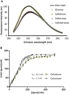
Figure 2. CebE of S. scabies interacts with cellobiose and cellotriose in the nanomolar range.
(A) Fluorescence emission spectra of the pure 6His-CebE protein (180 nM) alone (line) or in presence of 30 μM glucose (red circles), cellobiose (yellow circles), cellotriose (green circles) or cellotetraose (white circles). (B) CebE-sugar dissociation curves as a function of the cellobiose or cellotriose concentration. The fluorescence emission intensity of 180 nM 6His-CebE was measured in presence of different concentrations of cellobiose or cellotriose (from 15 to 500 nM). Each dissociation experiment was repeated 6 times. See materials and methods for details.
Figure 3
Figure 3. Effect of cebE and msiK deletion in S. scabies 87–22 on the production of thaxtomin.
(A) Growth and thaxtomin production (yellow pigmentation) on plates. Xylose and cellobiose were both supplied at 1% final quantity (weight/volume percentage concentration). (B) HPLC analysis of thaxtomin extracted from plates. Deletion of cebE or msiK in S. scabies resulted in the loss of the cellobiose mediated induction of thaxtomin production.
Figure 4
Figure 4. Effect of cebE and msiK deletion on the virulence of S. scabies.
Phenotypes of representative radish seedlings are shown after 7 days of growth treated with water, the wild type strains 87–22, and the cebE deletion mutant (A) or the msiK deletion mutant (B).
Figure 5
Figure 5. CebR directly represses transcription of genes encoding components of the cellobiose/cellotriose-specific ABC transporter.
(A,B) Effect of cebR deletion in S. scabies on the transcription levels of cebE, cebF, and msiK when grown on ISP-4 (A) and OBA (B). qPCR analysis of gene expression levels in S. scabies 87–22 and in the ΔcebR strain. Data were normalized using the gyrA and murX genes as internal controls. Mean normalized expression levels (±standard deviations) from three biological repeats analyzed in triplicate are shown. *Denotes significant quantitative overexpression (p > 0.05) in the S. scabies cebR null mutant compared to the wild-type strain 87–22. (C) CebR binds to the DNA motifs identified in the intergenic region between cebR and cebE (cbscebR-E) and upstream of bglC (cbsbglC) (see Fig. 1A for sequences and localization of cbs). EMSAs with increasing concentrations of pure CebR-His6, i.e., 0 30, 80, 160, 240, 320, 400, and 480 nM, respectively (numbers 1 to 8). (D) EMSAs demonstrating that cellobiose and cellotriose are able to inhibit the DNA-binding ability of CebR. Numbers 1 and 2 refer to EMSAs with free probes (6 nM) and with probes incubated with CebR-His6, respectively. Numbers 3 to 7 refer to EMSAs with CebR-His6 preincubated with oligosaccharides, i.e., cellobiose (lane 3), cellotriose (lane 4), cellotetraose (lane 5), cellopentaose (lane 6), or cellohexaose (lane 7).
Figure 6
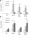
Figure 6
Relative abundancy of CebE (A) and MsiK (B) peptides in response to the deletion of cebR and/or cellobiose supply, determined by targeted proteomics (LC-MRM). Target peptides for CebE: ESDYLPWK (CebE1), SAFDLTAK (CebE2), and SGNWGGSFLSVPK (CebE3). Target peptides for MsiK: ILDLTEYLDR (MsiK1), TQIASLQR (MsiK2), and FGNSVVPVNR (MsiK3). *Denotes significant quantitative peptide overproduction (P < 0.05) compared to the wild-type strain grown in ISP-4 without cellobiose supply. Statistical significance was assigned by performing 2-sided Student’s t-tests and assuming groups of equal variances (See Supplementary Fig. S7).
Figure 7
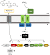
Figure 7. Model of the cellobiose/cellotriose-mediated induction of thaxtomin production in S. scabies.
Cellobiose (Glc)2 and Cellotriose (Glc)3 are transported by the CebEFG-MsiK ABC transporter. In the cytoplasm, (Glc)2 and, to a lesser extend (Glc)3, binds to CebR and inhibits its DNA-binding ability which further increases transcription of cebE, cebF, and cebG, as well as txtR, the specific activator of the thaxtomin A biosynthetic genes txtA and txtB. Circles (○) indicate the CebR-binding sites.

References

    1. Hopwood D. A. Streptomyces in Nature and Medicine: The Antibiotic Makers. (Oxford University Press, 2007).
    1. Hodgson D. A. Primary metabolism and its control in streptomycetes: a most unusual group of bacteria. Adv. Microb. Physiol. 42, 47–238 (2000). - PubMed
    1. Bertram R. et al.. In silico and transcriptional analysis of carbohydrate uptake systems of Streptomyces coelicolor A3(2). J. Bacteriol. 186, 1362–73 (2004). - PMC - PubMed
    1. Francis I., Holsters M. & Vereecke D. The Gram-positive side of plant-microbe interactions. Environ. Microbiol. 12, 1–12 (2010). - PubMed
    1. Kinkel L. L., Schlatter D. C., Bakker M. G. & Arenz B. E. Streptomyces competition and co-evolution in relation to plant disease suppression. Res. Microbiol. 163, 490–499 (2012). - PubMed

Publication types

MeSH terms