Review
. 2016 Jun 1;80(3):523-43.
doi: 10.1128/MMBR.00069-15. Print 2016 Sep.

Genetically Engineered Phages: a Review of Advances over the Last Decade

Affiliations
Review

Genetically Engineered Phages: a Review of Advances over the Last Decade

Diana P Pires et al. Microbiol Mol Biol Rev. .

Abstract

Soon after their discovery in the early 20th century, bacteriophages were recognized to have great potential as antimicrobial agents, a potential that has yet to be fully realized. The nascent field of phage therapy was adversely affected by inadequately controlled trials and the discovery of antibiotics. Although the study of phages as anti-infective agents slowed, phages played an important role in the development of molecular biology. In recent years, the increase in multidrug-resistant bacteria has renewed interest in the use of phages as antimicrobial agents. With the wide array of possibilities offered by genetic engineering, these bacterial viruses are being modified to precisely control and detect bacteria and to serve as new sources of antibacterials. In applications that go beyond their antimicrobial activity, phages are also being developed as vehicles for drug delivery and vaccines, as well as for the assembly of new materials. This review highlights advances in techniques used to engineer phages for all of these purposes and discusses existing challenges and opportunities for future work.


Figures

FIG 1
FIG 1
Phage engineering via homologous recombination. Upon phage infection (A), the injected DNA recombines with plasmid DNA carrying regions of homology (loci in red) to the phage genome (B), resulting in new, recombinant phage particles (fragments in orange) (C).
FIG 2
FIG 2
Bacteriophage recombineering of electroporated DNA. Purified phage DNA (A) and dsDNA recombineering substrates (B) are coelectroporated into cells (C). Recombination between their homologous regions (in orange) (D) results in recombinant phage particles (containing DNA fragments in green) (E).
FIG 3
FIG 3
In vivo recombineering. Bacterial cells carrying a defective λ prophage and the pL operon under the control of a temperature-sensitive repressor (in red) (A) are infected with the phage to be manipulated (B) and subsequently transformed with dsDNA or ssDNA (C). Recombination then occurs between the phage genome and the dsDNA/ssDNA (homologous loci in blue) (D), after which recombinant phage particles (carrying the DNA fragments in pink) are recovered (E).
FIG 4
FIG 4
CRISPR-Cas-mediated phage engineering. Upon phage infection, homologous recombination occurs between phage DNA (A) and plasmid DNA (B), such that a phage gene (in orange) is deleted. The resulting phage population is mixed (phages containing fragments in blue or orange) (C), but by using the CRISPR-Cas system (single guide RNA [sgRNA] is shown in orange and Cas proteins in red and yellow, encoded on separate plasmids) to target the gene retained in the wild-type particles (D), it is possible to counterselect the wild-type phage population (fragment in orange in genome) (E) and to retain the recombinant version (phage containing the blue-colored fragment) (F).
FIG 5
FIG 5
Rebuilding/refactoring of phage genomes in vitro. Once the phage DNA has been purified (A), it is digested using native restriction sites (B), and independent pieces (in burgundy) (C) can be subcloned and further manipulated (represented by the DNA fragments in burgundy, blue, and pink) (D). Once released from the vector, the recombinant section is ligated to the rest of the phage genome (E) and electroporated into the phage host for recovery of engineered phage particles (F).
FIG 6
FIG 6
Synthesis and assembly of phage genomes from synthetic oligonucleotides. Synthetic oligonucleotides (A) are annealed and assembled by PCA, followed by ligation (B). E. coli is subsequently transformed with the full circular genome molecules (C), and phage particles are recovered (D).
FIG 7
FIG 7
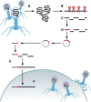
Yeast-based assembly of phage genomes. Purified phage DNA (A) is electroporated into S. cerevisiae together with linear YAC molecules with overhangs (in black) homologous to the 5′ and 3′ ends of the linear phage genome (B). Recombination in the yeast cell enables genomic subcloning (YAC backbone in green) (C), which upon YAC purification and electroporation (D) allows the recovery of phage particles (E).
FIG 8
FIG 8
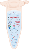
Cell-free systems for assembly of recombinant phage particles. Purified phage genome DNA is combined with cell-free expression systems (A) that enable gene transcription (B), translation (C), DNA replication (D), and assembly of whole phage particles (E).

References

    1. Twort FW. 1915. An investigation on the nature of ultra-microscopic viruses. Lancet 186:1241–1243. doi:10.1016/S0140-6736(01)20383-3. - DOI - PMC - PubMed
    1. d'Hérelle F. 1917. Sur un microbe invisible antagoniste des bacilles dysentériques. C R Hebd Seances Acad Sci Ser D 165:373–375.
    1. Bruynoghe R, Maisin J. 1921. Essais de thérapeutique au moyen du bacteriophage. C R Soc Biol 85:1120–1121.
    1. d'Hérelle F. 1921. Le bactériophage: son rôle dans l'immunité. Masson et Cie, Paris, France.
    1. Wittebole X, De Roock S, Opal SM. 2014. A historical overview of bacteriophage therapy as an alternative to antibiotics for the treatment of bacterial pathogens. Virulence 5:226–235. doi:10.4161/viru.25991. - DOI - PMC - PubMed

Publication types