

Plasticity of Candida albicans Biofilms
- PMID: 27250770
- PMCID: PMC4981664
- DOI: 10.1128/MMBR.00068-15
Plasticity of Candida albicans Biofilms
Abstract
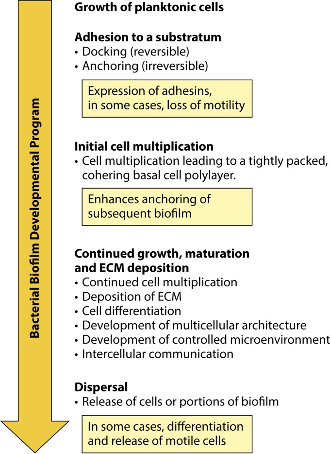
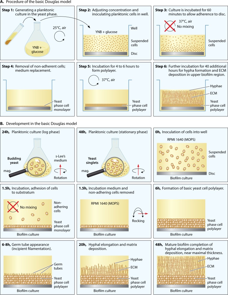
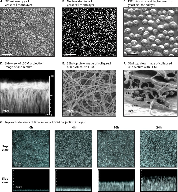
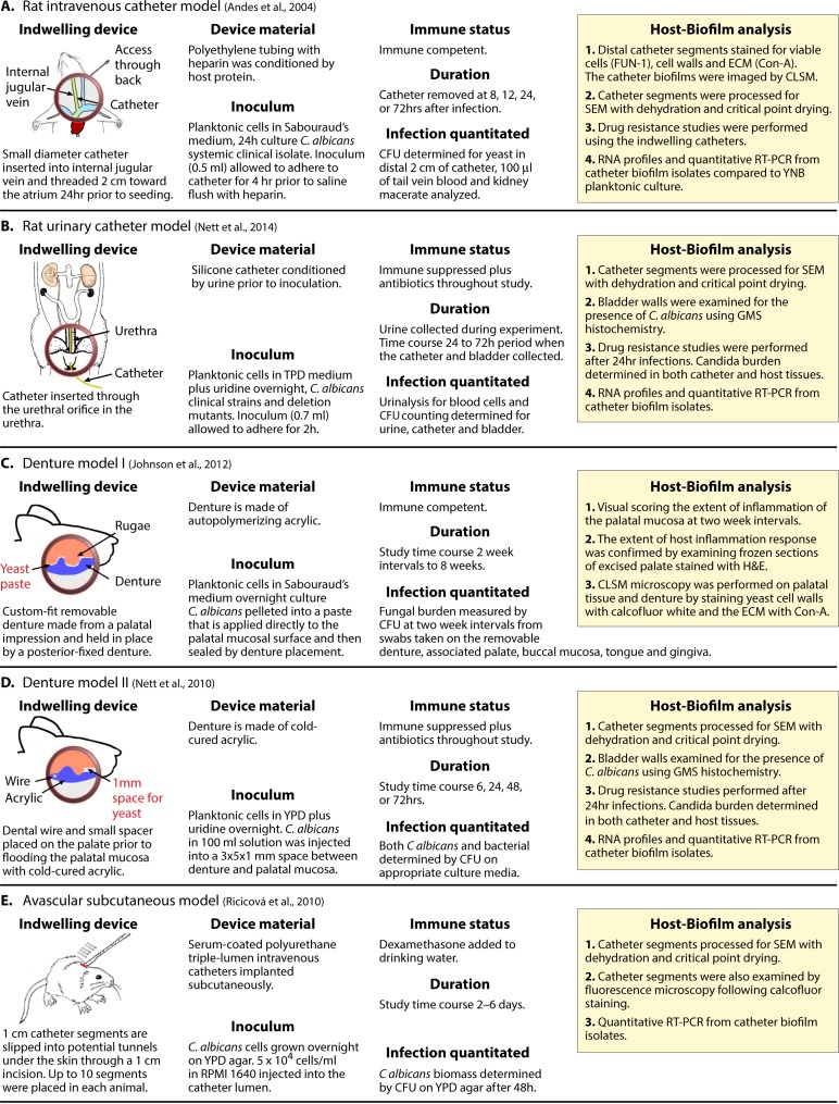
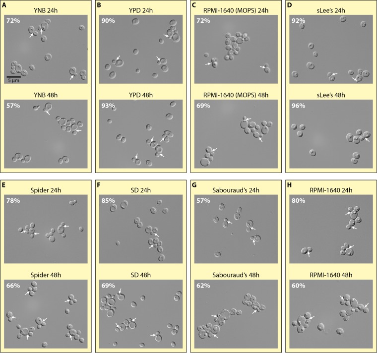
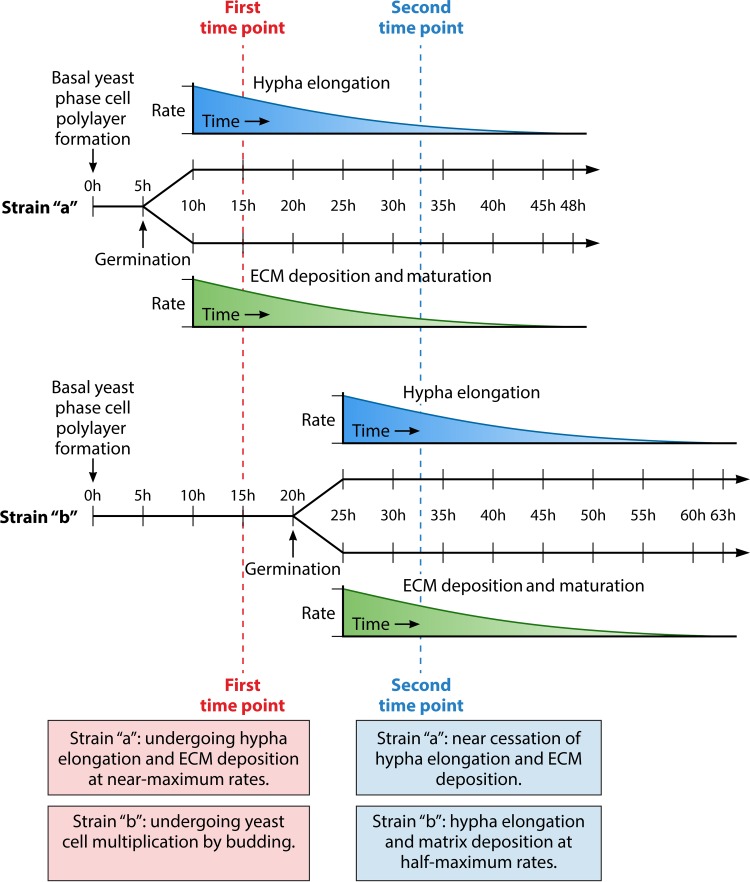
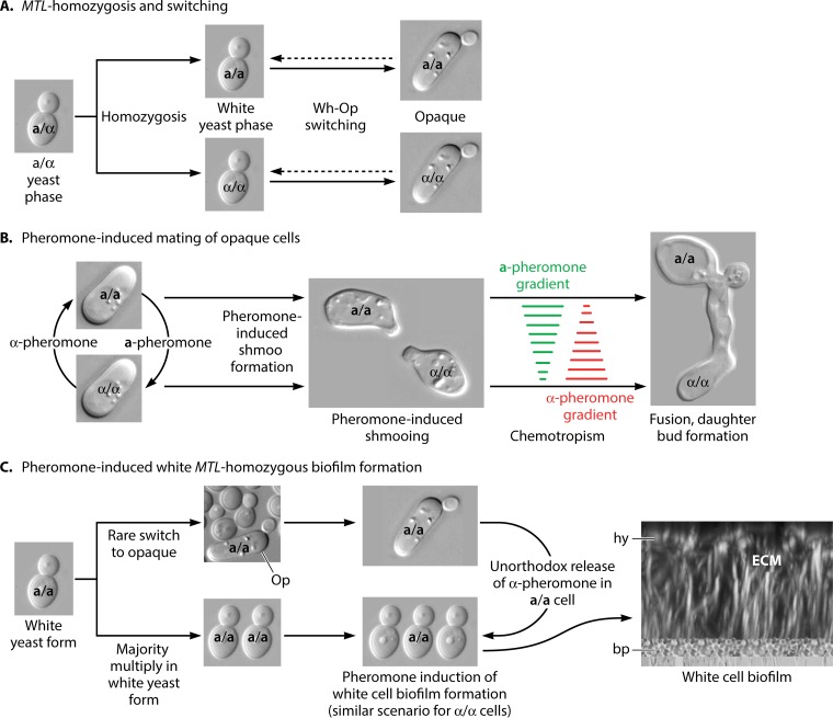
Candida albicans, the most pervasive fungal pathogen that colonizes humans, forms biofilms that are architecturally complex. They consist of a basal yeast cell polylayer and an upper region of hyphae encapsulated in extracellular matrix. However, biofilms formed in vitro vary as a result of the different conditions employed in models, the methods used to assess biofilm formation, strain differences, and, in a most dramatic fashion, the configuration of the mating type locus (MTL). Therefore, integrating data from different studies can lead to problems of interpretation if such variability is not taken into account. Here we review the conditions and factors that cause biofilm variation, with the goal of engendering awareness that more attention must be paid to the strains employed, the methods used to assess biofilm development, every aspect of the model employed, and the configuration of the MTL locus. We end by posing a set of questions that may be asked in comparing the results of different studies and developing protocols for new ones. This review should engender the notion that not all biofilms are created equal.
Copyright © 2016, American Society for Microbiology. All Rights Reserved.
Figures
Similar articles
-
Towards non-invasive monitoring of pathogen-host interactions during Candida albicans biofilm formation using in vivo bioluminescence.Cell Microbiol. 2014 Jan;16(1):115-30. doi: 10.1111/cmi.12184. Epub 2013 Sep 12. Cell Microbiol. 2014. PMID: 23962311 Free PMC article.
-
A rabbit model for evaluation of catheter-associated fungal biofilms.Virulence. 2011 Sep-Oct;2(5):466-74. doi: 10.4161/viru.2.5.16341. Epub 2011 Sep 1. Virulence. 2011. PMID: 21921676 Free PMC article.
-
Sustained Nitric Oxide-Releasing Nanoparticles Induce Cell Death in Candida albicans Yeast and Hyphal Cells, Preventing Biofilm Formation In Vitro and in a Rodent Central Venous Catheter Model.Antimicrob Agents Chemother. 2016 Mar 25;60(4):2185-94. doi: 10.1128/AAC.02659-15. Print 2016 Apr. Antimicrob Agents Chemother. 2016. PMID: 26810653 Free PMC article.
-
Candida albicans Biofilms and Human Disease.Annu Rev Microbiol. 2015;69:71-92. doi: 10.1146/annurev-micro-091014-104330. Annu Rev Microbiol. 2015. PMID: 26488273 Free PMC article. Review.
-
Portrait of Candida Species Biofilm Regulatory Network Genes.Trends Microbiol. 2017 Jan;25(1):62-75. doi: 10.1016/j.tim.2016.09.004. Epub 2016 Oct 4. Trends Microbiol. 2017. PMID: 27717660 Review.
Cited by
-
Screening of Candida albicans GRACE library revealed a unique pattern of biofilm formation under repression of the essential gene ILS1.Sci Rep. 2019 Jun 24;9(1):9187. doi: 10.1038/s41598-019-45624-y. Sci Rep. 2019. PMID: 31235750 Free PMC article.
-
Optimization of Candida tropicalis growth conditions on silicone elastomer material by response surface methodology.Front Bioeng Biotechnol. 2025 Jul 17;13:1572694. doi: 10.3389/fbioe.2025.1572694. eCollection 2025. Front Bioeng Biotechnol. 2025. PMID: 40746591 Free PMC article.
-
Secretions from Serratia marcescens Inhibit the Growth and Biofilm Formation of Candida spp. and Cryptococcus neoformans.J Microbiol. 2023 Feb;61(2):221-232. doi: 10.1007/s12275-022-00007-3. Epub 2023 Feb 21. J Microbiol. 2023. PMID: 36809632
-
Constitutive ALS3 expression in Candida albicans enhances adhesion and biofilm formation of efg1, but not cph1 mutant strains.PLoS One. 2023 Jul 13;18(7):e0286547. doi: 10.1371/journal.pone.0286547. eCollection 2023. PLoS One. 2023. PMID: 37440498 Free PMC article.
-
Automated quantification of Candida albicans biofilm-related phenotypes reveals additive contributions to biofilm production.NPJ Biofilms Microbiomes. 2020 Oct 9;6(1):36. doi: 10.1038/s41522-020-00149-5. NPJ Biofilms Microbiomes. 2020. PMID: 33037223 Free PMC article.
References
-
- Shapiro JA. 1988. Bacteria as multicellular organisms. Sci Am 258:82–89. - PubMed
Publication types
MeSH terms
LinkOut - more resources
Full Text Sources
Other Literature Sources
Medical
