

doi: 10.1101/pdb.prot091298.
Long-Term Potentiation by Theta-Burst Stimulation Using Extracellular Field Potential Recordings in Acute Hippocampal Slices
Affiliations
- PMID: 27250947
- PMCID: PMC5291724
- DOI: 10.1101/pdb.prot091298
Item in Clipboard
Long-Term Potentiation by Theta-Burst Stimulation Using Extracellular Field Potential Recordings in Acute Hippocampal Slices
Cold Spring Harb Protoc.
.
Abstract
This protocol describes how to carry out theta-burst long-term potentiation (LTP) with extracellular field recordings in acute rodent hippocampal slices. This method is relatively simple and noninvasive and provides a way to sample many neurons simultaneously, making it suitable for applications requiring higher throughput than whole-cell recording.
© 2016 Cold Spring Harbor Laboratory Press.
Figures
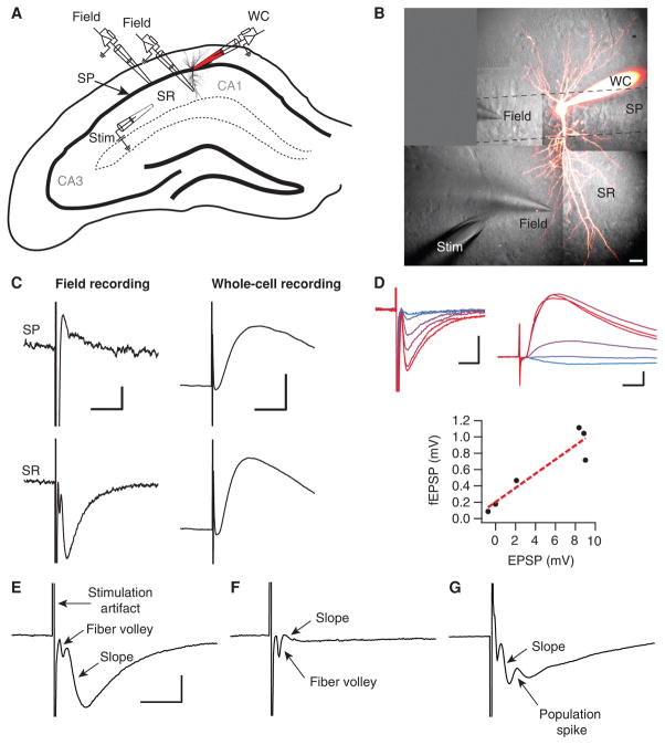
Factors determining fEPSP recordings. (A) Schematic drawing of a hippocampal slice showing positioning of stimulation and recording electrodes. A stimulation electrode (“stim”) is placed in the stratum radiatum (SR) of the CA1 area, to activate axons originating from pyramidal cells in CA3. For comparison, a whole-cell recording electrode (WC) is placed in the cell body layer, stratum pyramidale (SP). The field-recording electrode (“field”) is moved between SP and SR positions. (B) 2-photon microscopy maximal intensity projection recorded pyramidal cell loaded with Alexa 594 is superimposed on infrared Dodt contrast images. Positions of stimulation and field-recording electrodes are shown, similar to the schematic in A (scale bar: 20 μm). (C) fEPSPs recorded in SP (top left) and SR (bottom left) have reversed polarities (scale bars: 200 μV, 10 ms). EPSP recorded in whole-cell configuration remains the same (right, scale bars: 2 mV, 10 ms). Note that the SR fEPSP (bottom left) has faster dynamics and the opposite sign compared with the intracellular EPSP (bottom right). (D) fEPSPs (top left) and whole-cell EPSPs (top right) recorded simultaneously at six different stimulation intensities (30, 40, 50, 70, 80, and 100 V, indicated by lines ranging from blue to red) are significantly correlated (bottom; Pearson’s r = 0.933, P < 0.05), indicating equivalency in terms of measuring synaptic strength (scale bars: 500 μV, 5 ms [top left]; 2 mV, 5 ms [top right]). (E) Example of a good fEPSP recording. The stimulus artifact, fiber volley, and slope are indicated. The fEPSP is large compared with the fiber volley, which indicates a healthy slice (scale bar: 500 μV, 5 msec). (F) A fEPSP that is much smaller than the fiber volley suggests that the slice is of poor quality; despite stimulating many axons, the response is very small. (G) A fEPSP that has a population spike (positive deflection) riding on the PSP interfering with the measurement of the peak. This occurs when the recorded neurons fire.
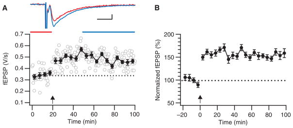
Theta-burst LTP. (A) Sample theta-burst induction experiment showing robust LTP. During the induction (arrow), 12 trains of 4 pulses at 100 Hz were delivered at 5 Hz, and this was repeated three times at 0.1 Hz. Error bars indicate xxx. Inset top: fEPSPs averaged over indicated time periods before (red) and after (blue) induction (scale bars: 100 μV, 5 ms). (B) This ensemble average of five such theta-burst experiments indicates how robust this LTP paradigm is. Before averaging, experiments were normalized to their individual baseline periods, so error bars (S.E.M.) reflect the variability around the mean, not absolute amplitude variability across experiments.
References
-
- Abrahamsson T, Gustafsson B, Hanse E. AMPA silencing is a prerequisite for developmental long-term potentiation in the hippocampal CA1 region. J Neurophysiol. 2008;100:2605–2614. - PubMed
-
- Artola A, Bröcher S, Singer W. Different voltage-dependent thresholds for inducing long-term depression and long-term potentiation in slices of rat visual cortex. Nature. 1990;347:69–72. - PubMed
Publication types
MeSH terms
Grants and funding
LinkOut - more resources
Full Text Sources
Other Literature Sources
Medical