. 2016 Jun 2;534(7605):129-32.
doi: 10.1038/nature17960. Epub 2016 May 25.

Overcoming EGFR(T790M) and EGFR(C797S) resistance with mutant-selective allosteric inhibitors

Affiliations

Overcoming EGFR(T790M) and EGFR(C797S) resistance with mutant-selective allosteric inhibitors

Yong Jia et al. Nature. .

Abstract

The epidermal growth factor receptor (EGFR)-directed tyrosine kinase inhibitors (TKIs) gefitinib, erlotinib and afatinib are approved treatments for non-small cell lung cancers harbouring activating mutations in the EGFR kinase, but resistance arises rapidly, most frequently owing to the secondary T790M mutation within the ATP site of the receptor. Recently developed mutant-selective irreversible inhibitors are highly active against the T790M mutant, but their efficacy can be compromised by acquired mutation of C797, the cysteine residue with which they form a key covalent bond. All current EGFR TKIs target the ATP-site of the kinase, highlighting the need for therapeutic agents with alternative mechanisms of action. Here we describe the rational discovery of EAI045, an allosteric inhibitor that targets selected drug-resistant EGFR mutants but spares the wild-type receptor. The crystal structure shows that the compound binds an allosteric site created by the displacement of the regulatory C-helix in an inactive conformation of the kinase. The compound inhibits L858R/T790M-mutant EGFR with low-nanomolar potency in biochemical assays. However, as a single agent it is not effective in blocking EGFR-driven proliferation in cells owing to differential potency on the two subunits of the dimeric receptor, which interact in an asymmetric manner in the active state. We observe marked synergy of EAI045 with cetuximab, an antibody therapeutic that blocks EGFR dimerization, rendering the kinase uniformly susceptible to the allosteric agent. EAI045 in combination with cetuximab is effective in mouse models of lung cancer driven by EGFR(L858R/T790M) and by EGFR(L858R/T790M/C797S), a mutant that is resistant to all currently available EGFR TKIs. More generally, our findings illustrate the utility of purposefully targeting allosteric sites to obtain mutant-selective inhibitors.


Figures

Extended Data Figure 1
Extended Data Figure 1. Inhibition of wild type and mutant EGFR kinases by EAI001 and EAI045 in purified enzyme assays
a, Inhibition of wild type and mutant EGFR kinases by EAI001. Activity of the indicated mutant EGFR kinase (residues 696-1022) was measured in the presence of increasing concentrations of EAI001. The HTRF assay was carried out using either 1 μM ATP (left panel) or 1 mM ATP (right panel). Error bars indicate standard deviation (n=2). b, Inhibition of L858R/T790M EGFR by EAI045 (left panel) or erlotinib (right panel) at a range of ATP concentrations, as indicated. Assay was performed using an HTRF-based assay as described in the Methods section of the main text. Error bars indicate standard deviation (n=2).
Extended Data Figure 2
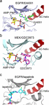
Extended Data Figure 2. Comparison of the binding site of EGFR allosteric inhibitors with those of lapatinib and allosteric MEK inhibitors
a, Structure of EAI001 in complex with EGFR for comparison. b, Structure of MEK1 kinase bound to allosteric inhibitor GDC0973 (PDB 4AN2). GDC0973 (also called XL518) and other allosteric MEK inhibitors occupy a pocket created by displacement of the C-helix in the inactive conformation of the kinase. Most allosteric MEK inhibitors make hydrogen bond interactions with the γ-phosphate group of ATP that are important for their potency. The allosteric EGFR inhibitors we describe here bind in a generally analogous location in EGFR, but lack any clear structural similarity to MEK inhibitors and do not contact the γ-phosphate group of ATP. c, The structure of lapatinib bound to EGFR (PDB ID 1XKK). Both lapatinib and neratinib (see Fig. 1d in the main text) bind an inactive conformation of the kinase. Like gefitinib and erlotinib, both occupy the ATP site, but also extend into the allosteric pocket occupied by EAI001. Note that like neratinib, lapatinib places aromatic phenyl or pyridinyl groups in positions similar to those occupied by the aminothiazole and phenyl substituents of EAI001.
Extended Data Figure 3
Extended Data Figure 3. EAI001 binding is incompatible with the inactive conformation of WT EGFR
Superposition of the EAI001/EGFR structure reported here with the structure of WT EGFR kinase in the inactive conformation (gray, PDB ID 2GS7). EAI001 (shown with carbon atoms in green) clashes with the side chains of leucines 858 and 861 in the WT EGFR structure. These leucine residues lie in a short helical segment at the N-terminus of the activation loop. The L858R substitution disrupts this helix. We propose that this effect explains, in part, the selectivity of the allosteric inhibitor for the L858R/T790M mutant. Note that EAI001 was crystallized with the T790M/V948R mutant EGFR, as we were unable to obtain crystals with the L858R/T790M or L858R/T790M/V948R proteins. The compound induces unstructuring of the activation loop helix and repositions L858, which is in contact with the 1-oxoisoindolinyl group of the inhibitor. The location and conformation of the inhibitor is expected to be the same in the context of the L858R mutation, but the details of the interaction with this portion of the activation loop will necessarily differ due to the mutation.
Extended Data Figure 4
Extended Data Figure 4. Cellular activity of EAI045
a, EAI045 inhibition of L858R/T790M EGFR in NIH-3T3 cells. Western blotting with the indicated concentrations of the allosteric inhibitor or with 1 μM WZ4002 as control (WZ) was carried out 6 h after compound addition. b-e, Profiling of EAI045 in Ba/F3 models bearing mutant EGFR or the parental Ba/F3 cell line, as indicted. Inhibition by WZ4002 is shown as a positive control. For gel source data, see Supplementary Figure 1.
Extended Data Figure 5
Extended Data Figure 5. Cellular and in vivo efficacy of EAI045 in combination with cetuximab
a, Ba/F3 cells bearing L858R/T790M/C797S EGFR were treated with EAI045 alone or with EAI045 plus cetuximab and proliferation was measured using the MTS assay after 72 hours. b, MRI imaging of cohorts L858R/T790M, exon19del/T790M, and L858R/T790M/C797S genetically engineered EGFR-mutant mice before treatment and 1 or 2 weeks after treatment with EAI045 and cetuximab. These cohorts of tumor bearing mice were used for short term efficacy and pharmacodynamic studies, and are distinct from those used for the tumor volume measurements shown in Fig. 3 in the main text.
Figure 1
Figure 1. Structure and binding mode of allosteric EGFR inhibitors
a, Chemical structures of EAI001 and EAI045. b, Overall view of the structure of T790M/V948R EGFR bound to EAI001 and AMP-PNP. EAI001 is shown in CPK form with carbon atoms in green. The V948R substitution was introduced to allow crystallization of the kinase in an inactive conformation. c, Detailed view of the interactions of EAI001. A hydrogen bond with Asp855 in the “DFG” segment of the kinase is shown as a dashed red line. d, The structure of irreversible inhibitor neratinib bound to EGFR T790M (PDB ID 2JIV). Neratinib occupies the ATP site, but also extends into the allosteric pocket occupied by EAI001.
Figure 2
Figure 2. Cellular activity and mechanism of synergy of EAI045 with cetuximab
a, Analysis of EAI045 inhibition of EGFR phosphorylation in H1975 cells by western blotting (anti-pY1068). A dose response study is shown at 3h after compound addition for EAI045 and the irreversible quinazoline inhibitor afatinib (control). For gel source data, see Supplementary Figure 1. b, The effect of EAI045 on EGFR target modulation in H1975 cells in the presence and absence of EGF. EGFR phosphorylation (pY1173) was measured using an ELISA-based assay; error bars indicate standard deviation (n=3). c, The allosteric pocket is differentially accessible in the two subunits of the asymmetric dimer. Unlike wild type EGFR in which only the receiver subunit is active, both subunits are catalytically active in the L858R/T790M mutant. The activator subunit is more readily inhibited by allosteric agents (yellow star), because the C-helix can be readily displaced. By contrast, opening the allosteric pocket in the receiver subunit requires perturbing the dimer. Thus mutations that disrupt the asymmetric dimer (such as I941R, blue circle) or antibodies that block dimerization (cetuximab) should enhance the potency of allosteric agents. d, Inhibition of proliferation of Ba/F3 cells expressing L858R/T790M and L858R/T790M/I941R by EAI045. Addition of the dimer-disrupting I941R mutation markedly sensitizes to inhibition by EAI045. e and f, Treatment of EGFR-Mutant Ba/F3 cells with EAI045 alone, in combination with cetuximab (10 μg/ml), or with cetuximab alone. Note the dramatic synergy with cetuximab that is observed only in the L858R/T790M model.
Figure 3
Figure 3. EAI045 in combination with cetuximab induces tumor regression in genetically engineered mouse models of EGFR-mutant lung cancer
a, Mice bearing L858R/T790M mutant tumors were treated with EAI045 alone (n=5), cetuximab alone (n=3) or both agents in combination (n=10). Tumor volumes were measured using MRI 4 weeks after initiation of treatment and are plotted for each animal in a “waterfall” format. b, As in panel a, but in mice bearing exon19del/T790M mutant tumors (n=4, 4 and 4). c, As in panel a, but in mice bearing L858R/T790M/C797S mutant tumors (n=3, 4, and 5). d and e, Pharmacodynamic studies in exon19del/T790M and L858R/T790M/C797S mice. Tumor nodules from mice treated with EAI045 or cetuximab alone or with the combination were analyzed by western blotting with the indicated antibodies to examine the effect of treatment on EGFR signaling. Multiple independent mouse tumors were obtained and analyzed, two independent and representative samples are shown. For gel source data, see Supplementary Figure 1. Source data for tumor volume measurements are provided in Supplementary Figure 2.

Comment in

References

    1. Mok TS, et al. Gefitinib or carboplatin-paclitaxel in pulmonary adenocarcinoma. N Engl J Med. 2009;361:947–957. - PubMed
    1. Yu HA, Pao W. Targeted therapies: Afatinib--new therapy option for EGFR-mutant lung cancer. Nature Reviews Clinical Oncology. 2013;10:551–552. - PMC - PubMed
    1. Gainor JF, Shaw AT. Emerging paradigms in the development of resistance to tyrosine kinase inhibitors in lung cancer. J Clin Oncol. 2013;31:3987–3996. - PMC - PubMed
    1. Chong CR, Janne PA. The quest to overcome resistance to EGFR-targeted therapies in cancer. Nature medicine. 2013;19:1389–1400. - PMC - PubMed
    1. Walter AO, et al. Discovery of a mutant-selective covalent inhibitor of EGFR that overcomes T790M-mediated resistance in NSCLC. Cancer Discov. 2013;3:1404–1415. - PMC - PubMed

Publication types

MeSH terms