Case Reports
. 2016 Oct;43(10):884-91.
doi: 10.1111/cup.12746. Epub 2016 Jun 29.

Distinct genetic profiles of extracranial and intracranial acral melanoma metastases

Affiliations
Case Reports

Distinct genetic profiles of extracranial and intracranial acral melanoma metastases

Gaurav Sharma et al. J Cutan Pathol. 2016 Oct.

Abstract

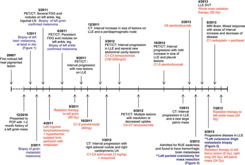
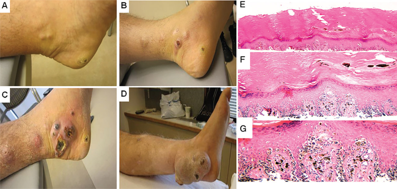
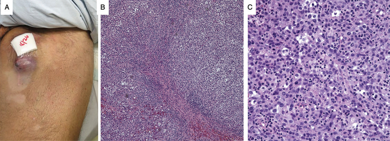
Background: There is limited knowledge of the genetic alterations in acral melanoma metastases at different anatomic sites. Here, we characterized the genetic abnormalities of metastases in a 51-year-old man with stage IIIC heel melanoma who developed concomitant brain and cutaneous metastases in spite of multiple treatment modalities.

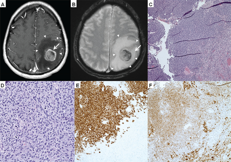
Methods: Melanoma cells were isolated following palliative resection of the patient's cortical tumor and biopsy of cutaneous thigh metastasis. Mutational analysis using polymerase chain reaction amplification and BLAST, as well as exome sequencing (160 Mb coverage) was performed on the tumors, cell lines generated thereof and normal lymph nodes.

Results: All specimens had neuroblastoma RAS viral oncogene homolog Q61K mutations. There was a 40-fold higher somatic mutation frequency in the brain metastasis compared to the cutaneous metastasis. The former showed truncations of DNA mismatch repair genes (MLH1 and MSH2), and non-canonical BRAF (v-raf murine sarcoma viral oncogene homolog B1), PIK3CA and NF-1 mutations not observed in the extracranial lesion. Genomic profiling of each cell line was concordant with the respective original tumor tissue.

Conclusions: We present the mutational differences between brain and cutaneous acral melanoma metastases in a patient with concomitant lesions. Further genetic and functional studies are needed to understand the biology of metastatic disease appearing at different sites.

Keywords: NRAS; acral melanoma; brain metastases; cutaneous metastases; mismatch repair genes.


Figures

Fig. 1.
Fig. 1.
Timeline of clinical course, pathological specimen (highlighted in blue) and treatments (highlighted in red). PCP, primary care physician; PET/CT, positron emission tomography/computed tomography; FDG, fluorodeoxyglucose; LN, lymph node; Gy, gray; LLE, left lower extremity; C, cycle; RUE, right upper extremity.
Fig. 2.
Fig. 2.
Primary acral melanoma. A–D) Clinical photos of the lesion. A) s/p biopsy of heel primary melanoma (March 2011), (B) positron emission tomography shows interval increase in lesions in left lower extremity (LLE) (December 2011), (C) interval progression on LLE while on ipilimumab (March 2012) and (D) persistent lesion s/p radiation therapy (July 2013). E–G) Histology of the lesion. E) Lower power view of superficial shave biopsy of acral melanoma, (F) medium power view shows atypical melanocytes confluent along the basal layer and (G) high power view exhibits pagetoid spread, and the severe atypia of the melanocytes. There is minimal dermis in the specimen; however, no definitive invasion is seen.
Fig. 3.
Fig. 3.
Left cutaneous thigh metastasis. A) Gross photograph of left cutaneous thigh metastasis, (B) low and C) high power view of the H&E from the cutaneous thigh metastasis shows sheets of atypical melanocytes with nuclear pleomorphism and several mitoses.
Fig. 4.
Fig. 4.
Intracranial metastasis. Magnetic resonance imaging in (A) axial T1 and (B) T2-weighted imaging showed an enhancing 3-cm lesion (arrows) with areas of hemorrhage. The mass involved the motor cortex with marked, surrounding vasogenic edema (arrowheads) and local mass effect manifested as sulcal effacement. The histopathology shown on H&E with a (C) low power image, (D) high power image and positive immmunostains (E) Mart-1 and (F) S100 exhibits diffuse melanoma.

References

    1. Hodis E, Watson IR, Kryukov GV, et al. A landscape of driver mutations in melanoma. Cell 2012; 150: 251. - PMC - PubMed
    1. Lawrence MS, Stojanov P, Polak P, et al. Mutational heterogeneity in cancer and the search for new cancer-associated genes. Nature 2013; 499: 214. - PMC - PubMed
    1. Long GV, Stroyakovskiy D, Gogas H, et al. Combined BRAF and MEK inhibition versus BRAF inhibition alone in melanoma. N Engl J Med 2014; 371: 1877. - PubMed
    1. Larkin J, Chiarion-Sileni V, Gonzalez R, et al. Combined nivolumab and ipilimumab or monotherapy in untreated melanoma. N Engl J Med 2015; 373: 23. - PMC - PubMed
    1. Berger MF, Levin JZ, Vijayendran K, et al. Integrative analysis of the melanoma transcriptome. Genome Res 2010; 20: 413. - PMC - PubMed

Publication types

MeSH terms