. 2016 Jul 26;87(4):410-8.
doi: 10.1212/WNL.0000000000002827. Epub 2016 Jun 1.

A new saccadic indicator of peripheral vestibular function based on the video head impulse test

Affiliations

A new saccadic indicator of peripheral vestibular function based on the video head impulse test

Hamish G MacDougall et al. Neurology. .

Abstract

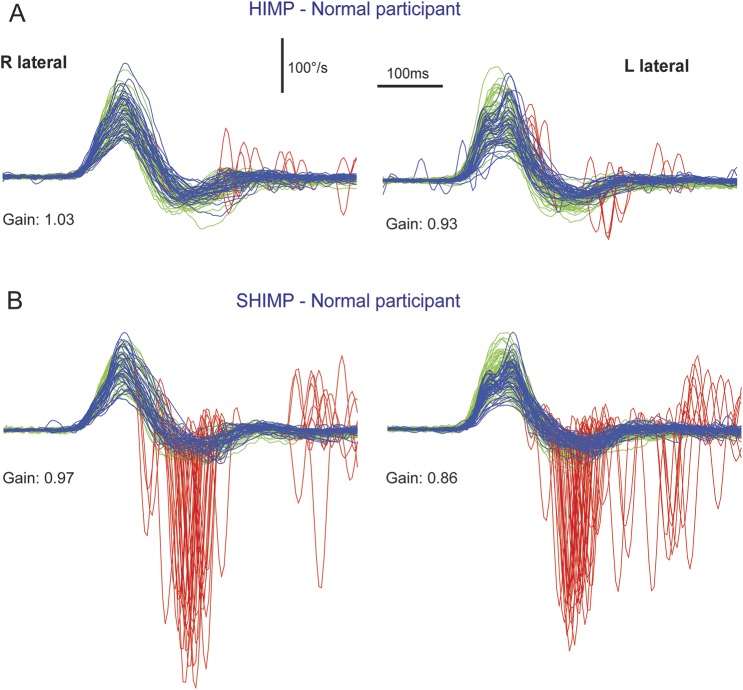
Objective: While compensatory saccades indicate vestibular loss in the conventional head impulse test paradigm (HIMP), in which the participant fixates an earth-fixed target, we investigated a complementary suppression head impulse paradigm (SHIMP), in which the participant is fixating a head-fixed target to elicit anticompensatory saccades as a sign of vestibular function.

Methods: HIMP and SHIMP eye movement responses were measured with the horizontal video head impulse test in patients with unilateral vestibular loss, patients with bilateral vestibular loss, and in healthy controls.

Results: Vestibulo-ocular reflex gains showed close correlation (R(2) = 0.97) with slightly lower SHIMP than HIMP gains (mean gain difference 0.06 ± 0.05 SD, p < 0.001). However, the 2 paradigms produced complementary catch-up saccade patterns: HIMP elicited compensatory saccades in patients but rarely in controls, whereas SHIMP elicited large anticompensatory saccades in controls, but smaller or no saccades in bilateral vestibular loss. Unilateral vestibular loss produced covert saccades in HIMP, but later and smaller saccades in SHIMP toward the affected side. Cumulative HIMP and SHIMP saccade amplitude differentiated patients from controls with high sensitivity and specificity.

Conclusions: While compensatory saccades indicate vestibular loss in conventional HIMP, anticompensatory saccades in SHIMP using a head-fixed target indicate vestibular function. SHIMP saccades usually appear later than HIMP saccades, therefore being more salient to the naked eye and facilitating vestibulo-ocular reflex gain measurements. The new paradigm is intuitive and easy to explain to patients, and the SHIMP results complement those from the standard video head impulse test.

Classification of evidence: This case-control study provides Class III evidence that SHIMP accurately identifies patients with unilateral or bilateral vestibulopathies.


Figures

Figure 1
Figure 1. Video head impulse test of a healthy control with SHIMP compared to conventional HIMP
During SHIMP (B), the participant's task is to fixate a target, which is moving with the head, whereas in conventional HIMP (A), the target remains stationary. The figure illustrates the typical HIMP and SHIMP saccade pattern in a healthy control. (A) During conventional HIMP, a healthy control elicits only few mostly positive catch-up saccades (red) after the end of the head impulse. (B) During SHIMP, the same healthy control shows large negative saccades after the end of the head impulse reflecting anticompensatory eye movements back to the head-fixed target. Both paradigms give similar but slightly lower vestibulo-ocular reflex gain values during SHIMP compared to HIMP, but a complementary saccade pattern. Head velocity = green traces; inverted slow phase eye velocity = blue traces; saccades = red traces; HIMP = conventional head impulse paradigm; SHIMP = suppression head impulse paradigm.
Figure 2
Figure 2. Video head impulse test of a patient with BVL using SHIMP compared to conventional HIMP
Typical patient with complete BVL showing a reversed saccadic pattern during HIMP and SHIMP compared to a healthy control (figure 1). (A) During standard HIMP, the patient with BVL elicits mostly overt positive catch-up saccades after the head impulse. (B) During SHIMP, the same patient with BVL shows only very few downward saccades reflecting anticompensatory saccades after the end of the head impulse back to the head-fixed target. Both paradigms give similar but slightly lower vestibulo-ocular reflex gain values during SHIMP compared to HIMP, but a complementary saccade pattern, which is reversed compared to healthy controls. Head velocity = green traces; inverted slow phase eye velocity = blue traces; saccades = red traces. BVL = bilateral vestibular loss; HIMP = conventional head impulse paradigm; SHIMP = suppression head impulse paradigm.
Figure 3
Figure 3. Video head impulse test of a patient with UVL using SHIMP compared to conventional HIMP
Typical patient with UVL showing reversed saccadic patterns during HIMP compared to SHIMP to the healthy and affected side. (A, affected) With standard HIMP, the patient elicits stereotyped covert saccades during head impulses to the affected side. (B, affected) With SHIMP, the patient elicits only small negative saccades after impulses to the affected side. Note that compared to HIMP (A, affected), SHIMP (B, affected) clears the eye velocity traces from covert saccades during head impulses to the affected side, thus facilitating gain calculation. Head impulses to the healthy side produce only small negative saccades during HIMP (A, healthy), but large negative saccades during SHIMP (B, healthy). Vestibulo-ocular reflex gain values to the healthy side are slightly lower during SHIMP compared to HIMP, but very similar to the affected right side. Head velocity = green traces; inverted slow phase eye velocity = blue traces; saccades = red traces; HIMP = conventional head impulse paradigm; SHIMP = suppression head impulse paradigm; UVL = unilateral vestibular loss.
Figure 4
Figure 4. Cumulative saccade amplitude as a function of latency after head impulse onset
(A) In healthy controls, HIMP elicits only a few saccades (upward histogram bars), while SHIMP elicits a multitude of saccades (downward histogram bars) with a peak latency of about 176 milliseconds (ms). (C) Patients with BVL show a reversed saccadic pattern with large saccades in HIMP but only a few saccades in SHIMP. Patients with UVL often produce covert HIMP saccades with head impulses to the affected side (B) and overt SHIMP saccades to the healthy side (D). Note that in the same patients with UVL, overt SHIMP saccades to the healthy side (D) have a longer peak latency (176 ms) compared to the covert HIMP saccades to the affected side (104 ms, B). Histogram bars represent summated amplitudes of HIMP saccades (positive) and SHIMP saccades (negative) in 8-ms bins after head impulse onset. Saccade amplitude was normalized relative to the number of head impulses and participants and kept in proportion between participant groups (A, n = 6 controls × 2 sides), patients with UVL (B, affected side; D, healthy side, n = 5), and patients with BVL (C, n = 5 × 2). BVL = bilateral vestibular loss; HIMP = conventional head impulse paradigm; SHIMP = suppression head impulse paradigm; UVL = unilateral vestibular loss.
Figure 5
Figure 5. Video head impulse test model for illustration of saccade size in relation to VOR gain deficit
(A) In a healthy control, a head impulse with an earth-fixed target (HIMP) elicits no saccade. (B) In the same healthy control with a VOR gain of one, a head impulse with a head-fixed target (SHIMP) elicits an anticompensatory saccade of the size of the head rotation (16.5°). (J) Conversely, in a patient with total bilateral vestibular loss with a VOR gain of zero, SHIMP elicits no saccade while HIMP elicits a saccade the size of the head rotation (I). (C) Little VOR loss (gain 0.9) is sufficient to elicit a small compensatory saccade with HIMP. (H) In a patient with incomplete vestibular loss, little residual function (gain 0.1) is sufficient to elicit a small anticompensatory saccade with SHIMP. Note that on visual inspection in the velocity domain, the amplitude of smaller saccades (C, 1.5° amplitude) is overestimated compared to the amplitude of larger saccades (I, 15.8° amplitude). HIMP = conventional head impulse paradigm; SHIMP = suppression head impulse paradigm; VOR = vestibulo-ocular reflex.

References

    1. Halmagyi GM, Curthoys IS. A clinical sign of canal paresis. Arch Neurol 1988;45:737–739. - PubMed
    1. MacDougall HG, McGarvie LA, Halmagyi GM, Curthoys IS, Weber KP. The video head impulse test (vHIT) detects vertical semicircular canal dysfunction. PLoS One 2013;8:e61488. - PMC - PubMed
    1. MacDougall HG, Weber KP, McGarvie LA, Halmagyi GM, Curthoys IS. The video head impulse test: diagnostic accuracy in peripheral vestibulopathy. Neurology 2009;73:1134–1141. - PMC - PubMed
    1. Weber KP, Aw ST, Todd MJ, McGarvie LA, Curthoys IS, Halmagyi GM. Head impulse test in unilateral vestibular loss: vestibulo-ocular reflex and catch-up saccades. Neurology 2008;70:454–463. - PubMed
    1. Barin K. Interpretation and usefulness of caloric testing. In: Jacobson GP, Shepard NT, editors. Balance Function Assessment and Management, 2nd ed. San Diego: Plural Publishing; 2016:319–345.

Publication types