Review
. 2016 Jun 1;8(6):a005918.
doi: 10.1101/cshperspect.a005918.

Protein Regulation in Signal Transduction

Affiliations
Review

Protein Regulation in Signal Transduction

Michael J Lee et al. Cold Spring Harb Perspect Biol. .

Abstract

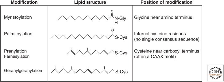
SUMMARYCells must respond to a diverse, complex, and ever-changing mix of signals, using a fairly limited set of parts. Changes in protein level, protein localization, protein activity, and protein-protein interactions are critical aspects of signal transduction, allowing cells to respond highly specifically to a nearly limitless set of cues and also to vary the sensitivity, duration, and dynamics of the response. Signal-dependent changes in levels of gene expression and protein synthesis play an important role in regulation of protein levels, whereas posttranslational modifications of proteins regulate their degradation, localization, and functional interactions. Protein ubiquitylation, for example, can direct proteins to the proteasome for degradation or provide a signal that regulates their interactions and/or location within the cell. Similarly, protein phosphorylation by specific kinases is a key mechanism for augmenting protein activity and relaying signals to other proteins that possess domains that recognize the phosphorylated residues.


Figures

Figure 1.
Figure 1.
Allosteric modulation of protein activity. (A) Cartoon example of protein allostery. In this example, an allosteric-regulation site exists in a region of the protein that is spatially distinct from the active site. Modulation of the allosteric site (through posttranslational modification or the binding of a ligand, cofactor, or protein) causes changes in the active site. Importantly, these changes can be either activating or inhibiting. (B) Allosteric regulation of dihydrofolate reductase (DHFR). (Left panels) Two surface views of the DHFR enzyme, highlighting the active site (bound to folate), cofactor binding site (bound to NADPH), and residues involved in allosteric communication between the two sites (shown in blue). (Middle panels) A view of a slice through the protein core. (Right panels) A cartoon representation of the slice mappings shown in B. (Figure generously supplied by Rama Ranganathan and Cell Press.)
Figure 2.
Figure 2.
Mechanism of kinase activation. (A) Conformational changes in protein kinases upon phosphorylation enhance their catalytic ability. The structure of ERK2, an MAPK, is shown in its inactive nonphosphorylated state and its active phosphorylated state. The carboxy-terminal lobes of the kinase in both states (brown and red, respectively) have been superimposed. Note that following phosphorylation of the activation loop, there is a marked rotation and reorientation of the amino-terminal lobe (yellow and purple, respectively), bringing key catalytic residues, including those present in a critical α helix, αC, into position, converting the kinase into an active state that can now phosphorylate downstream substrates. (B) Close-up of phosphorylation-induced conformational changes in the activation loop. Two key residues in the activation loop of MAPKs, a threonine and a tyrosine residue, separated by a singe amino acid (i.e., a TXY motif) can interact with a network of surrounding arginine residues only when they are in their phosphorylated states. These interactions not only shift the positions of the threonine and tyrosine residues themselves (curved arrows), but drag the entire activation loop into a new conformation that communicates with the rest of the protein to move the entire amino-terminal lobe relative to the carboxy-terminal lobe, as shown in A.
Figure 3.
Figure 3.
Examples of protein posttranslational modifications and modular protein-binding domains that recognize these modified amino acids. (A) Structures of common amino acid posttranslational modifications. The parent amino acid structure is shown in black; the modification is shown in red. (B) Cartoon representations of modular binding domains. α Helices are shown in cyan; β-strands are shown in purple; loops are shown in orange. SH2 domains recognize peptides containing phosphotyrosine, FHA domains recognize peptides containing phosphothreonine, Bromo domains recognize peptides containing acetyl-lysine, and Tandem Tudor domains recognize dimethylarginine. In the examples shown, the SH2 domain is from Src kinase, the FHA domain is from Chk2, the Bromo domain is from Brd4, and the Tandem Tudor domains are from SND1.
Figure 4.
Figure 4.
The GTPase cycle. G proteins can be small (Ras-like) or large (heterotrimeric). Depicted here is the nucleotide cycle for heterotrimeric G proteins, but the reactions are conceptually the same for small GTPases. G-protein-coupled receptors (GPCRs) bind extracellular ligands, and transmit signals to intracellular G proteins. The ligand-bound receptor functions as a guanine-nucleotide exchange factor (GEF), causing the Gα subunit to exchange GDP for GTP. GTP-bound Gα no longer interacts with the Gβγ dimer, and both entities are free to interact with downstream effector proteins. Gα controls the duration of the signal because it is a GTPase, whose activity can be stimulated by GTPase-activating proteins (GAPs) such as RGS proteins.
Figure 5.
Figure 5.
A multistep mechanism for maximal Src activation. (A) In the inactive state, Src is folded up as a consequence of multiple interactions between the reader domains and motifs in Src itself. The Src SH2 domain is bound to a phosphotyrosine residue (Y527) in the carboxyl terminus, while the SH3 domain binds to a polyproline-type helix in the linker that connects the SH2 domain to the kinase domain. (B) The kinase opens up into an active conformation when a ligand such as a growth factor receptor or an adaptor protein engages the SH2 and SH3 domains directly, usually accompanied by dephosphorylation of the Y527 site to prevent intramolecular reassociation into the closed form. (C) Autophosphorylation of Y416 in the activation loop of Src, or phosphoprylation of this site by another kinase, results in maximal activity. (From Xu et al. 1999; adapted, with permission, © Elsevier.)
Figure 6.
Figure 6.
Common forms of protein lipidation. The table highlights four lipid moieties that are commonly used to modify proteins posttranslationally or (in the case of myristoylation) cotranslationally. These modifications differ in terms of their consensus sequences, position of the modification, hydrophobicity, and mechanism of regulation.
Figure 7.
Figure 7.
Protein ubiquitylation. (A) Structure of the ubiquitin monomer, highlighting the amino and carboxyl termini, as well as key lysine residues. (B) Schematic depiction of the posttranslational modification of substrate proteins with ubiquitin. Ubiquitin is added to proteins through a three-step enzymatic reaction featuring a ubiquitin-activating enzyme (E1), a ubiquin-conjugating enzyme (E2), and a ubiquitin ligase (E3). Subsequent rounds of ubiquitylation can result in the formation of ubiquitin chains. (C) Ubiquitin chain diversity. Ubiquitin contains seven lysine residues, each of which can be used as the anchorage point for subsequent ubiquitin monomers, in homotypic (same linkage throughout chain), heterotypic (mixed chains), or branched fashion (multiple ubiquitin monomers conjugated to a single ubiquitin). In addition, the amino-terminal methionine on a ubiquitin monomer linked to a substrate protein can be linked to the carboxy-terminal end of another ubiquin monomer (linear chains). Although all of these forms have been shown to exist in cells, physiological roles for many of these chains are still being elucidated.

References

    1. Alexander J, Lim D, Joughin BA, Hegemann B, Hutchins JRA, Ehrenberger T, Ivins F, Sessa F, Hudecz O, Nigg EA, et al. 2011. Spatial exclusivity combined with positive and negative selection of phosphorylation motifs is the basis for context-dependent mitotic signaling. Sci Signal 4: ra42. - PMC - PubMed
    1. Ambros V. 2001. microRNAs: Tiny regulators with great potential. Cell 107: 823–826. - PubMed
    1. Ambros V. 2011. MicroRNAs and developmental timing. Curr Opin Genet Dev 21: 511–517. - PMC - PubMed
    1. Azeloglu EU, Iyengar R. 2014. Signaling networks: Information flow, computation, and decision making. Cold Spring Harb Perspect Med 10.1101/cshperspect.a005934. - DOI - PMC - PubMed
    1. Bieging KT, Attardi LD. 2012. Deconstructing p53 transcriptional networks in tumor suppression. Trends Cell Biol 22: 97–106. - PMC - PubMed

LinkOut - more resources