. 2016 Jul 27;90(16):7248-7256.
doi: 10.1128/JVI.00542-16. Print 2016 Aug 15.

Mutagenesis of S-Adenosyl-l-Methionine-Binding Residues in Coronavirus nsp14 N7-Methyltransferase Demonstrates Differing Requirements for Genome Translation and Resistance to Innate Immunity

Affiliations

Mutagenesis of S-Adenosyl-l-Methionine-Binding Residues in Coronavirus nsp14 N7-Methyltransferase Demonstrates Differing Requirements for Genome Translation and Resistance to Innate Immunity

James Brett Case et al. J Virol. .

Abstract

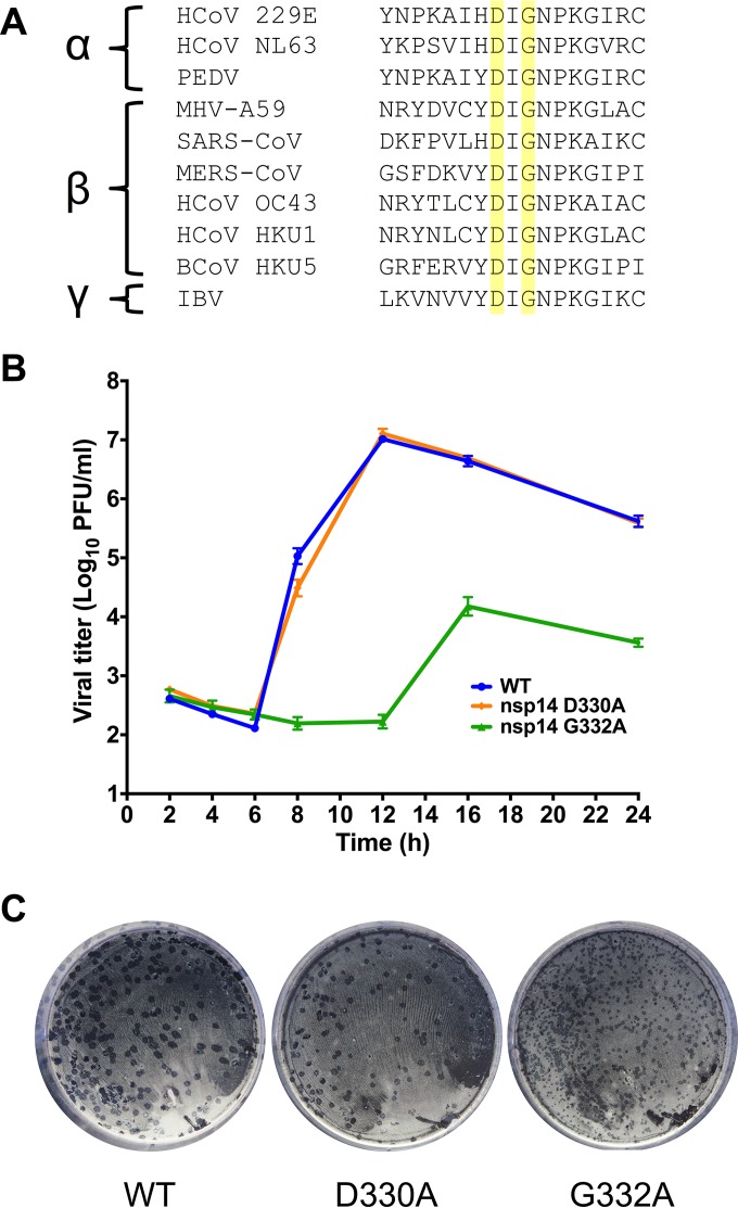
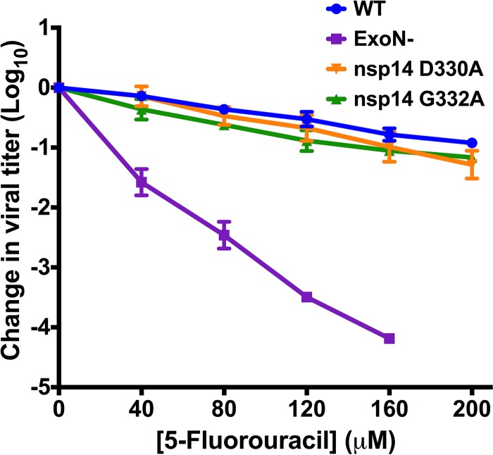
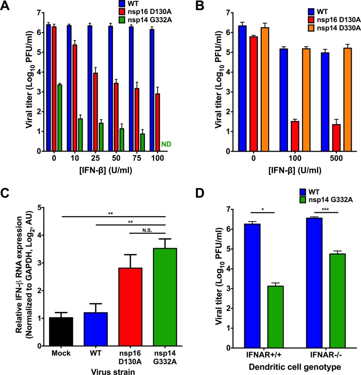
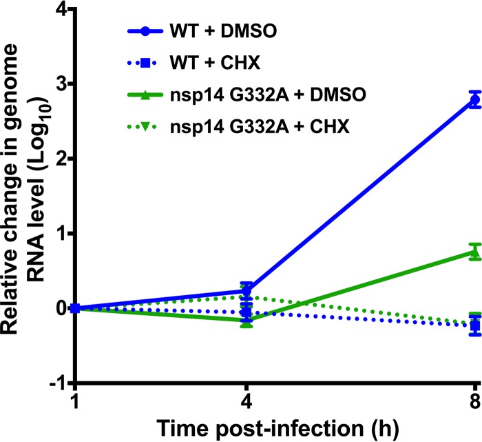
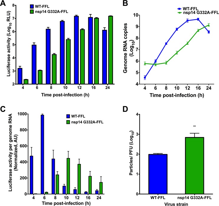
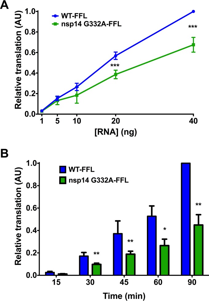
Eukaryotic mRNAs possess a methylated 5'-guanosine cap that is required for RNA stability, efficient translation, and protection from cell-intrinsic defenses. Many viruses use 5' caps or other mechanisms to mimic a cap structure to limit detection of viral RNAs by intracellular innate sensors and to direct efficient translation of viral proteins. The coronavirus (CoV) nonstructural protein 14 (nsp14) is a multifunctional protein with N7-methyltransferase (N7-MTase) activity. The highly conserved S-adenosyl-l-methionine (SAM)-binding residues of the DxG motif are required for nsp14 N7-MTase activity in vitro However, the requirement for CoV N7-MTase activity and the importance of the SAM-binding residues during viral replication have not been determined. Here, we engineered mutations in murine hepatitis virus (MHV) nsp14 N7-MTase at residues D330 and G332 and determined the effects of these mutations on viral replication, sensitivity to mutagen, inhibition by type I interferon (IFN), and translation efficiency. Virus encoding a G332A substitution in nsp14 displayed delayed replication kinetics and decreased peak titers relative to wild-type (WT) MHV. In addition, replication of nsp14 G332A virus was diminished following treatment of cells with IFN-β, and nsp14 G332A genomes were translated less efficiently both in vitro and during viral infection. In contrast, substitution of alanine at MHV nsp14 D330 did not affect viral replication, sensitivity to mutagen, or inhibition by IFN-β compared to WT MHV. Our results demonstrate that the conserved MHV N7-MTase SAM-binding-site residues are not required for MHV viability and suggest that the determinants of CoV N7-MTase activity differ in vitro and during virus infection.

Importance: Human coronaviruses, most notably severe acute respiratory syndrome (SARS)-CoV and Middle East respiratory syndrome (MERS)-CoV, cause severe and lethal human disease. Since specific antiviral therapies are not available for the treatment of human coronavirus infections, it is essential to understand the functions of conserved CoV proteins in viral replication. Here, we show that substitution of alanine at G332 in the N7-MTase domain of nsp14 impairs viral replication, enhances sensitivity to the innate immune response, and reduces viral RNA translation efficiency. Our data support the idea that coronavirus RNA capping could be targeted for development of antiviral therapeutics.


Figures

FIG 1
FIG 1
Replication kinetics of viruses with altered N7-MTase SAM-binding residues. (A) Alignment of GenBank ORF1b sequences of the α-, β-, and γ-CoVs shown demonstrates that SAM-binding residues (highlighted in yellow) are highly conserved. (B) DBT cells were infected with the viruses shown at an MOI of 1 PFU/cell. Cell culture supernatants were collected at the indicated times postinfection, and viral titers were determined by plaque assay. Error bars indicate standard errors of the means (SEM) (n = 6). (C) Plaque morphology of the viruses following agarose overlay plaque assay and fixation with 3.7% paraformaldehyde 24 h postinfection.
FIG 2
FIG 2
N7-MTase mutants display WT-like sensitivity to the RNA mutagen 5-FU. DBT cells were treated with the indicated concentrations of 5-FU for 30 min prior to infection with the viruses shown at an MOI of 0.01 PFU/cell. Medium containing 5-FU or vehicle was added 30 min postinfection. After 24 h, cell culture supernatants were collected, and viral titers were determined by plaque assay. For each virus, titers were normalized to those following infection of DMSO-treated controls. Changes in viral titers for nsp14 D330A and nsp14 G332A viruses were not statistically significant relative to WT MHV by one-way ANOVA. Error bars indicate SEM (n = 4).
FIG 3
FIG 3
nsp14 G332A virus exhibits increased induction of and sensitivity to IFN-β. DBT cells were treated for 18 h with the indicated concentrations of mouse IFN-β. Cells were infected with WT, nsp16 D130A, or nsp14 G332A virus and incubated for 24 h (A) or infected with WT, nsp16 D130A, and nsp14 D330A virus and incubated for 12 h (B). Cell culture supernatants were collected, and viral titers were determined by plaque assay. For each panel, error bars represent SEM (n = 4). ND, not detectable. (C) DBT cells were treated for 18 h with 10 U/ml mouse IFN-β. Cells were mock infected or infected with WT, nsp16 D130A, or nsp14 G332A virus at an MOI of 0.1 PFU/cell. At 12 h postinfection, cell lysates were harvested, total RNA was extracted, cDNA was generated, and IFN-β expression relative to GAPDH was determined by qPCR. Error bars indicate SEM (n = 9). N.S., not significant; **, P < 0.01 by Student's t test. (D) BMDCs were infected with either WT or nsp14 G332A virus at an MOI of 0.01 PFU/cell. At 24 h postinfection, cell culture supernatants were collected, and viral titers were determined by plaque assay. Error bars indicate SEM (n = 6). *, P < 0.05; ***, P < 0.001 by Student's t test.
FIG 4
FIG 4
nsp14 G332A genomic RNAs are stable. DBT cells were infected with WT or nsp14 G332A virus at an MOI of 0.01 PFU/cell in the presence of vehicle (DMSO) or 100 μg/ml CHX. Cell lysates were harvested at the indicated times postinfection and spiked with a known amount of in vitro-transcribed Renilla luciferase RNA, and total RNA was obtained by phenol-chloroform extraction. cDNA was generated by RT-PCR, and the number of viral genome copies present relative to Renilla luciferase was determined by SYBR green qPCR using MHV nsp10 and Renilla luciferase-specific primers. Error bars indicate SEM (n = 6).
FIG 5
FIG 5
nsp14 G332A genomic RNAs are translated with delayed kinetics during infection. DBT cells were infected with either WT-FFL or nsp14 G332A-FFL virus at an MOI of 0.1 PFU/cell. At the times shown postinfection, cell culture supernatants were collected, and lysates were harvested and divided equally into two samples. For the first lysate sample, luciferase activity was quantified (A). For the remaining lysate sample, RNA was extracted, and genome RNA copies were quantified using real-time qRT-PCR with a standard curve and CoV nsp2-specific primers (B). (C) Translation of WT-FFL or nsp14 G332A-FFL genomes at the times shown postinfection as determined by luciferase activity per genome RNA copy number. Values were normalized to WT-FFL at 6 h postinfection. Error bars indicate SEM (n = 4). (D) Viral titers in cell culture supernatants from DBT cells infected with either WT-FFL or nsp14 G332A-FFL were determined by plaque assay, and the number of genome RNA copies present in the input supernatant was determined by one-step real-time qRT-PCR. The particle-to-PFU ratio was calculated by dividing the number of genome RNA copies by the viral titers. Error bars represent SEM (n = 4). **, P < 0.01 by Student's t test.
FIG 6
FIG 6
Purified nsp14 G332A genomic RNA is translated at lower efficiency in vitro. BHK-R cells were infected at an MOI of 0.001 PFU/cell with either WT-FFL or nsp14 G332A-FFL virus. Supernatants were harvested and clarified, and virions were collected by ultracentrifugation. Virion pellets were resuspended, TRIzol was added, and virion RNAs were purified using phenol-chloroform phase separation. Genome RNA copies were quantified using one-step real-time qRT-PCR with a standard curve and CoV nsp2-specific primers. (A) The concentrations of WT-FFL or nsp14 G332A-FFL genomic RNAs shown were translated in vitro at 30°C for 1.5 h, and luciferase activity was quantified. Translation values are relative to WT-FFL genomic RNA at 40 ng. Error bars represent SEM (n = 4). ***, P < 0.001 by Student's t test. (B) Equivalent numbers of either WT-FFL or nsp14 G332A-FFL genomic RNAs were translated in vitro for the times shown, and luciferase activity was quantified. Error bars represent SEM (n = 6). *, P < 0.05; **, P < 0.01 by Student's t test.

References

    1. Shatkin AJ. 1976. Capping of eucaryotic mRNAs. Cell 9:645–653. doi: 10.1016/0092-8674(76)90128-8. - DOI - PubMed
    1. Darnell JE. 1979. Transcription units for mRNA production in eukaryotic cells and their DNA viruses. Prog Nucleic Acid Res Mol Biol 22:327–353. doi: 10.1016/S0079-6603(08)60803-X. - DOI - PubMed
    1. Furuichi Y, Shatkin AJ. 2000. Viral and cellular mRNA capping: past and prospects. Adv Virus Res 55:135–184. doi: 10.1016/S0065-3527(00)55003-9. - DOI - PMC - PubMed
    1. Marcotrigiano J, Gingras A-C, Sonenberg N, Burley SK. 1997. Cocrystal structure of the messenger RNA 5′ cap-binding protein (eIF4E) bound to 7-methyl-GDP. Cell 89:951–961. doi: 10.1016/S0092-8674(00)80280-9. - DOI - PubMed
    1. Filipowicz W, Furuichi Y, Sierra JM, Muthukrishnan S, Shatkin AJ, Ochoa S. 1976. A protein binding the methylated 5′-terminal sequence, m7GpppN, of eukaryotic messenger RNA. Proc Natl Acad Sci U S A 73:1559–1563. doi: 10.1073/pnas.73.5.1559. - DOI - PMC - PubMed

Publication types

MeSH terms